Skin Rashes

Chapter 43


Skin Rashes


Primary dermatologic diagnoses rarely require admission to the intensive care unit (ICU), but many patients in the ICU develop dermatologic manifestations of their underlying disease or as complications of therapy. Dermatologic disorders complicate many ICU stays and prolong length of stay. This chapter describes a variety of benign and serious skin problems relevant to ICU patients and their physicians.


Common Benign Skin Rashes (Table 43.1)
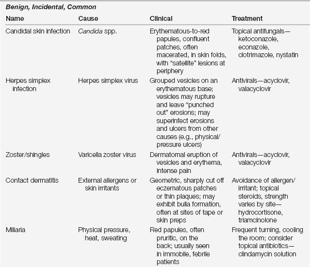


Candidal Infections


Candidiasis is most commonly caused by Candida albicans and occasionally by other candidal species. C. albicans, a yeast, inhabits the gastrointestinal tract, urinary tract, and intertriginous areas of the skin. The warmth and moisture of intertriginous areas provide a hospitable environment for growth. Higher skin pH, diapers, occlusive products, alteration of normal flora with systemic antibiotic use, and immunocompromised states all facilitate candidal overgrowth. C. albicans commonly infects the mucous membranes and skin, although in immunocompromised states there may be disseminated disease. In patients thus affected, untreated cutaneous candida infection may progress to widespread disease and even candidemia.


Candidal intertrigo occurs commonly and can involve any skin fold, especially in overweight individuals. Classically, the skin folds develop erythematous, beefy red patches, sometimes with an edge of macerated, superficially denuded skin. Frequently, tiny white pustules occur at the edges, and smaller red “satellite” lesions may be scattered just beyond the main rash. The skin lesions are painful when macerated and often pruritic.


When mucous membranes are involved, loosely adherent, creamy, well-demarcated curdlike debris on an erythematous base is typically seen on the tongue, buccal mucosa, gums, and palate (“thrush”). Oropharyngeal infection often progresses to involve the vermilion border at the angle of the mouth, causing maceration and fissuring known as perlèche. Severe mucosal candidal infection can extend to the esophagus and pharynx; esophageal candidiasis should prompt investigation of an underlying immunocompromised state—for example, as a presenting sign of human immunodeficiency virus (HIV) infection and representing an AIDS-defining illness. Treat oral candidiasis with anticandidal troches (clotrimazole), and intertriginous disease with topical creams (ketoconazole, econazole) twice daily. As intertrigo is frequently polymicrobial, topical therapy with soaks (acetic acid compresses), and keeping the area dry with barrier pastes (zinc oxide, triple paste) can be both therapeutic and preventive.


Contact Dermatitis


Contact dermatitis occurs from the interaction of a chemical with the skin. There are two major forms: irritant and allergic. Irritant contact dermatitis accounts for 80% of cases and results from damage to the skin via a toxic effect intrinsic to the compound; these irritants react with any skin and do not require prior sensitization. Common irritants in the ICU are soaps, stool, and urine, which when left in prolonged contact with the skin cause direct toxicity. These reactions typically occur within minutes to hours of the exposure, depending on the concentration of the irritant and the integrity of the skin.


Allergic contact dermatitis, a type IV cell-mediated delayed hypersensitivity immune response (see also Table 32.1 in Chapter 32), accounts for the other 20% of cases. It is antigen specific, and the reaction requires prior sensitization. In the ICU, patients may become sensitized to a variety of topical preparations, such as iodine, topical antimicrobials, or the adhesives used in tape or electrodes.


Generally, contact dermatitis appears as a well-demarcated, pruritic, erythematous, eczematous patch in a linear or geographic pattern corresponding to where the external agent was applied to the skin. Severe reactions can be vesicular or bullous in nature. Detection of the causative allergen can be determined by patch testing or by a “use test,” applying the agent in question several times daily for 5 to 7 days to an unaffected area of skin in an attempt to invoke the reaction. Treatment is avoidance of the causative allergen or irritant and midstrength topical steroids, such as triamcinolone ointment 0.1%, for mild cases and more potent topical steroids, such as clobetasol propionate ointment 0.05% or betamethasone dipropionate 0.05%, for severe reactions. Topical treatment is twice daily for 10 to 14 days. Rarely, systemic steroids are required for extensive severe contact allergy. Conditions that fail to respond to therapy should prompt consideration of alternate diagnoses.


Herpes Simplex Virus Skin Infections


Herpes simplex virus (HSV) is a common acquired infection caused by HSV type 1 or 2. Clinically, HSV appears as grouped vesicles on an erythematous base, especially in the perioral or genital areas. As the infection progresses, the vesicles may be lost, and the morphology instead consists of numerous “punched out” shallow erosions, often with a distinctive scalloped border. This acquired infection often recurs after trauma, ultraviolet light exposure, illness, or stress, as the virus infects nerves and lays dormant. Patients describe burning, itching, or stinging, which frequently precedes clinical lesions.


Diagnosis is made by Tzanck smear revealing multinucleated giant cells with nuclear molding, or by direct fluorescent antibody (DFA), polymerase chain reaction (PCR), or viral culture. In treatment-resistant cases, one should perform a viral culture to assess for drug resistance and guide therapy. Immunocompromised hosts may present with unusual morphologies and atypical lesions. HSV infection of the oral cavity may extend distally down the esophagus in intubated patients. HSV can occur at sites of pressure or trauma, such as around endotracheal or nasogastric tubes, and while erosions at those sites may be from physical causes, clinicians should consider whether viral superinfection may be present in atypical cases.


Topical treatment may reduce discomfort and promote healing; topical acyclovir, penciclovir, and tetracaine are variably efficacious. Oral acyclovir may decrease viral shedding, pain and crusting, and duration of disease if initiated early in the course of illness. In severe cases, such as most cases in the ICU or in immunocompromised patients, intravenous acyclovir may be used. Other antiviral options include famciclovir and valacyclovir. Resistant cases may require intravenous foscarnet or cidofovir.


Herpes Zoster


Herpes zoster, also known as shingles, is a vesicular eruption that occurs in a dermatomal distribution. Herpes zoster is caused by the reactivation of latent varicella zoster virus (VZV), which lays dormant in sensory neurons. Age and immunosuppression are the main risk factors for reactivation. The development of a zoster vaccine may eventually decrease cases in the elderly. Clinically, vesicles are seen on an erythematous base, distributed unilaterally in a dermatome, most commonly on the trunk (see Figure 101.1 in Chapter 101 for a discussion of sensory dermatomes). Occasionally the lesions become hemorrhagic; frequently they evolve to crusted erosions and then heal over the course of 2 to 3 weeks. Burning, stinging, itching, or pain often precedes the cutaneous manifestations. Up to 20 lesions can be found outside the affected dermatome, but a more extensive spread of lesions warrants prompt investigation for signs of disseminated varicella infection.


Initiate antiviral therapy as soon as possible, optimally within the first 72 hours of disease. Antiviral therapy promotes lesion healing and recovery, and it also decreases the risk of postzoster pain syndromes. Valacyclovir and famciclovir are preferable to acyclovir. Given that zoster is seen frequently in elderly patients, appropriate dosing for age-adjusted renal function is essential. Local therapy with heat pads, pressure, and support may provide additional relief; and gabapentin use may reduce the risk of post-zoster pain. Intravenous therapy is occasionally necessary for the immunocompromised patient and is always required for disseminated disease.
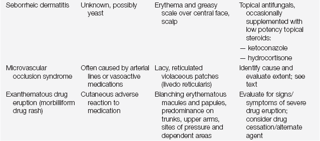


Miliaria


Miliaria results from obstruction of eccrine sweat ducts at various levels of the epidermis and dermis; as a result, sweat is retained in the skin. Miliaria crystalline manifests as asymptomatic, noninflammatory, small, fragile, superficial vesicles that rupture at the slightest contact. Miliaria rubra occurs as distinct, pruritic red papules and papulovesicles. Miliaria pustulosa consists of superficial, nonfolliculocentric pustules in intertriginous areas, on flexural surfaces, and on the back. These lesions may all appear after repeated episodes of fevers and sweating. Miliaria is fairly common in bed-confined ICU patients because of a combination of heat, moisture, and occlusion of the skin.


The cause of the obstruction is not clear. The condition resolves within days and treatment is symptomatic, keeping the area dry and unoccluded. Cooling the room or providing a fan for circulation, coupled with frequent turning and laundry changes, may help. Hydrophilic ointments or drying powders may also provide relief. Topical antibiotics, such as erythromycin or clindamycin, may also be used, as they may both dry out the skin and help to manage potentially causative microorganisms. Patients with severe pruritus may feel relief from short courses of mild- to moderate-strength topical steroids (triamcinolone ointment 0.1%).


Seborrheic Dermatitis


Seborrheic dermatitis is a common, benign papulosquamous disease. It affects sebaceous-rich areas such as the scalp, eyebrows, nasolabial folds, and central chest. Clinically, seborrheic dermatitis appears as erythematous macules and very thin plaques with greasy scale that can be pruritic.


The cause is unclear, but Pityrosporum ovale, a yeast found on normal skin and increased in seborrheic dermatitis, has been implicated. Seborrheic dermatitis flares in ill individuals and is associated with neurologic conditions such as Parkinson’s disease or strokes, which may lead to unilateral seborrheic dermatitis on the affected side. Patients with HIV not only have a higher incidence of the disease but also have manifestations that are more extensive, severe, and resistant to therapy. The disease is chronic with a waxing and waning course. Treatment is with topical antifungal agents, such as ketoconazole cream twice daily, short courses of low-potency steroids (hydrocortisone ointment 1% or 2.5%) as needed, and medicated shampoos (selenium sulfide or ketoconazole).


Drug Reactions


Patients in the ICU typically receive multiple medications, placing them at risk for developing adverse cutaneous drug reactions. These reactions can range from common and relatively benign morbilliform eruptions to more serious and even life-threatening reactions such as erythema multiforme major or Stevens-Johnson syndrome (SJS), toxic epidermal necrolysis (TEN), drug reaction with eosinophilia and systemic symptoms (DRESS), and drug-induced vasculitides. All cases of severe adverse drug reactions should be managed in conjunction with an experienced dermatology consultant.


Morbilliform Drug Rash


The most common drug reaction seen is a morbilliform eruption; this morphologic pattern comprises up to 90% of all cutaneous adverse drug reactions. Classically, patients present with erythematous macules and papules. Lesions may exhibit preference for dependent areas and sites of pressure or friction such as elastic bands, adhesives, areas of skin-on-skin rubbing, and the sacral region. Mucosal surfaces should be examined for involvement, which may portend a more serious drug reaction.


This reaction typically takes place 4 to 14 days after starting a new medication and is presumed to be a type IV hypersensitivity reaction, although the immunologic pathophysiology has not been completely elucidated. A thorough query of the patient’s medication history is essential to try to determine the most likely causative agent. Because it is difficult to identify the inciting agent accurately, discontinue any nonessential drugs. In limited eruptions with no concerning features (worrisome features include involvement of mucosal surfaces, skin pain, blistering, necrosis, or progression to involve sites typically spared, such as acral surfaces) some cases of morbilliform drug eruptions can be managed conservatively, and it may be possible to continue administering the causative agent if the implicated drug is essential to the patients’ care. In the majority of cases, however, it is better to discontinue the suspect medication and note the allergy in the patient’s medical record. One should perform daily cutaneous examinations to watch for progression. The drugs most frequently associated with morbilliform eruptions include allopurinol, aminopenicillins, cephalosporins, antiepileptic drugs, and antibacterial sulfonamides (see Chapter 32).


After discontinuing a suspected drug, the rash can worsen for a few days and persist for up to 2 weeks. Erythematous areas may exhibit superficial, nonpainful desquamation, resembling resolving sunburn. Symptomatic treatment of pruritus with topical midstrength steroids (triamcinolone ointment 0.1%) and lubricants (e.g., petroleum jelly, Absorbase) may be helpful.

< div class='tao-gold-member'>

Stay updated, free articles. Join our Telegram channel

Jul 7, 2016 | Posted by in CRITICAL CARE | Comments Off on Skin Rashes

Full access? Get Clinical Tree

Get Clinical Tree app for offline access