89 Hand and Wrist Injuries
 Key Points
Epidemiology
Annually, more than 16 million people suffer some form of hand injury, and more than 4.8 million will seek treatment of these injuries at the emergency department (ED).1 Traumatic injuries may include lacerations, fractures, and tendon or ligamentous injuries. Most injuries are readily identified in the ED, provided that the assessment is effective.2 Because the morbidity associated with undiagnosed, misdiagnosed, or mistreated hand injuries is significant, a high level of expertise is required of the EP.
Presenting Signs and Symptoms
During the history the patient’s hand dominance and career should also be ascertained and documented. Factors that may compromise wound healing, such as smoking, drug use, or an immunocompromised state, are important to document.2 Tetanus status should be verified.
Despite its complicated nature, the hand can be examined adequately in a short period. Developing a rapid, reproducible hand examination strategy and performing it regularly will decrease the chance of missing subtle injuries. Even when a specific injury is obvious, it is important to examine the entire hand to avoid overlooking less obvious, coincident injuries. Box 89.1 lists one approach to comprehensive assessment of the hand.
Box 89.1 Two-Minute Hand Examination
Ligamentous
Flexor digitorum profundus tendon—hold the proximal interphalangeal joint in extension; flex the distal interphalangeal joint against resistance
Flexor digitorum sublimis tendon—hold the metacarpophalangeal joint in extension; flex the proximal interphalangeal joint against resistance
Extensor tendons—place the hand palm down; extend the digit with resistance at the nail bed
Ulnar collateral ligament—adduct the thumb against resistance
Assuming that no active bleeding is occurring and requires immediate attention, examination of the hand begins with inspection. All rings, watches, and other potentially constricting devices should be removed immediately.3 Lacerations and other disruptions in skin integrity are usually recognized easily; erythema, soft tissue swelling, and ecchymoses should also be noted. It is important to compare the general position of the hand with that of the unaffected side inasmuch as many fractures or tendon disruptions will cause characteristic deformities that are recognizable on inspection.
Differential Diagnosis and Medical Decision Making
Fractures of the Hand
Metacarpal Bone Fractures
Metacarpal base fractures are uncommon and usually of little significance.2 The exception is at the base of the fifth metacarpal bone, where there can be associated subluxation of the metacarpal-hamate joint. The injured hand should be immobilized in an ulnar gutter splint and scheduled for referral to hand surgery.
Bennett/Reverse Bennett and Rolando Fractures
A Bennett fracture is a fracture of the proximal first metacarpal bone (Fig. 89.1). The classic mechanism is an axial load onto a flexed and adducted thumb. For example, a football quarterback strikes the helmet of an opposing player after releasing a throw. In this avulsion injury, the strong abductor pollicis longus muscle fractures the bone at the point of its insertion at the ulnar aspect of the first metacarpal bone. This causes displacement of a bony fragment, which can be seen on a plain film radiograph. It is usually an unstable fracture, and ED management should consist of referral to a hand surgeon and immobilization in a thumb spica splint. Potential long-term morbidity includes malunion, decreased function, and significant arthritis.
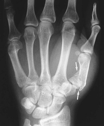
Fig. 89.1 Bennett fracture (arrows).
(From Mettler Jr FA. Essentials of radiology. 2nd ed. Philadelphia: Saunders; 2004.)
Boxer’s Fracture
Although many people refer to any fracture of the fifth metacarpal as a boxer’s fracture, the specific injury is a fracture through the neck of the bone (Fig. 89.2). This injury is most frequently seen when a solid object is forcefully struck with a closed fist. True boxer’s fractures may carry significant morbidity because in addition to being an unstable fracture, the injury often has a rotational component. If allowed to heal in this position, the hand will be deformed and weakened.
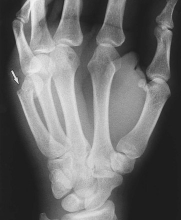
Fig. 89.2 Boxer’s fracture (arrow).
(From Mettler Jr FA. Essentials of radiology. 2nd ed. Philadelphia: Saunders; 2004.)
Distal Phalanx Fractures
The most common distal phalanx fracture is a tuft fracture, and nail bed injuries are the most common complication of this fracture.2 There is some controversy regarding the need to repair nail bed injuries; however, most authors recommend performing trephination only for nail bed hematomas involving 30% to 50% or greater of the nail bed surface. When the nail bed is involved, tuft fractures are considered open fractures. Although some physicians prescribe antibiotics empirically, evidence suggests that prophylactic antibiotics are not indicated.4 Proximal distal phalanx fractures are often unstable and require hand surgeon referral for percutaneous wire placement. An attempt to reduce any rotational deformity or angulation should be made before splinting. Splinting should isolate the DIP joint alone.


