Fig. 18.1
Evaluation of peptic ulcer disease (PUD)-associated benign GOO
The classically described test for GOO (i.e., “gastric retention”) is the saline load test described by Goldstein et al. in 1965 [24]. After evacuation of the stomach via a nasogastric tube (NGT), 750 mL of normal saline is instilled into the stomach over 3–5 min, and gastric contents aspirated 30 min later. In Goldstein’s study of 92 subjects, of whom 69 were controls, they found that greater than 400 mL of saline remaining in the stomach at 30 min was highly suggestive of gastric retention [24]. They further noted that reversion of the test to normal with medical treatment indicated that surgery would not be required for that episode of GOO [24].
Imaging
Plain films of the abdomen may reveal massive gastric distention with an air-fluid level in the stomach and the absence of small bowel distention [5, 10]. Additional imaging studies that can aid in the diagnosis include upper gastrointestinal (UGI) contrast studies and computerized tomography (CT) scans [9]. Barium radiography typically reveals three layers in the stomach: air, retained gastric juice, and sediments at the bottom [5]. In normal individuals, the majority of the barium slurry will be emptied from the stomach within 2 h and all of it by 6 h on UGI study, whereas in GOO, more than 60% of liquid barium will be retained in the stomach for more than 4 h and some of it can be retained for more than 24 h [9, 10]. In interpreting these studies, it should be recalled that the t1/2 of gastric emptying for water is around 10–20 min, depending on proximal gastric tone. Gastric emptying of solids is about 1–4 h and varies according to ease of liquefaction, contraction intensity, and composition of the meal [13]. Giant peristaltic waves may appear early during obstruction while a distended, atonic stomach during decompensated GOO may be noted on imaging studies [5]. Although the use of barium is commonly described, water-soluble contrast material is also used. Each contrast agent has advantages and disadvantages. CT scanning will further assist in the evaluation for malignant etiologies [9].
Gastric emptying scintigraphy was described by Griffith et al. in 1966 and has generally been regarded as the gold standard for evaluation of gastric emptying [25, 26]. Scintigraphy is generally performed for up to 2 h using 99mTc-sulfur colloid or 99mTc-DTPA [26]. A variety of test meal compositions (liquid, solid, or a combination thereof), as well as a variety of positions for testing, have been described [26]. Some reasons for the use of scintigraphy are its simplicity, reproducibility, and quantitative ability [27]. Of note, while several symptoms are often noted with GOO, a nuclear medicine study demonstrated that various commonly described symptoms of GOO such as epigastric discomfort, postprandial fullness, etc. are not reliable indicators of GOO or gastroparesis [27]. As such, objective testing for GOO is advisable.
Endoscopy
Endoscopy is the primary modality for localizing the site of obstruction and evaluating pathology [9]. GOO is generally diagnosed when a 9–11 mm endoscope cannot be passed through the stenosis [8, 9]. The degree of stenosis is assessed by comparing the size of the opening to the outer diameter of the endoscope and the ability to advance the endoscope into the duodenum, distal to the obstruction [28]. When performing endoscopy, biopsy is also advised to evaluate for malignancy and other, non-PUD-associated, causes of GOO [9]. Caution, however, must be exercised when solely using endoscopy to exclude malignancy in GOO, as a retrospective study of 40 patients with GOO found that endoscopic biopsy, including repeat biopsy and jumbo biopsy, had a sensitivity of only 37% for malignancy [29]. In a multivariate analysis, age and negative history of PUD were associated with increased risk for malignant GOO [29]. Given these findings, it was suggested that if the initial biopsy is negative then at least one more set of larger endoscopic biopsies should be performed in patients older than age 55 and without a history of PUD [29]. Even if the second set of biopsies is also negative for malignancy, a CT of the abdomen and pelvis for these high-risk individuals is still advisable [29].
Labs
Possible laboratory findings from persistent vomiting are a hypochloremic, hypokalemic metabolic alkalosis, secondary to loss of gastric contents that have high concentrations of sodium, chloride, and hydrogen [10, 13]. Additional chemistry findings include mild to moderate hyponatremia, increased serum bicarbonate, elevated BUN, and elevated creatinine [9]. A complete blood count may demonstrate hemoconcentration and normal or mildly elevated white blood cell count [9]. It may also demonstrate anemia; this may only become apparent with resuscitation [19]. A urinalysis may demonstrate high urine-specific gravity and paradoxically, aciduria [9]. The vomiting-induced loss of volume, sodium, and potassium forces the kidney to conserve sodium. To retain sodium, the kidney secretes hydrogen ions into the glomerular filtrate, resulting in paradoxical aciduria [9]. Potassium is also lost in the urine. The treatment is administration of isotonic saline to replace sodium and chloride deficits. Replacement of the sodium deficit in turn allows the kidney to excrete alkaline urine [5].
Management
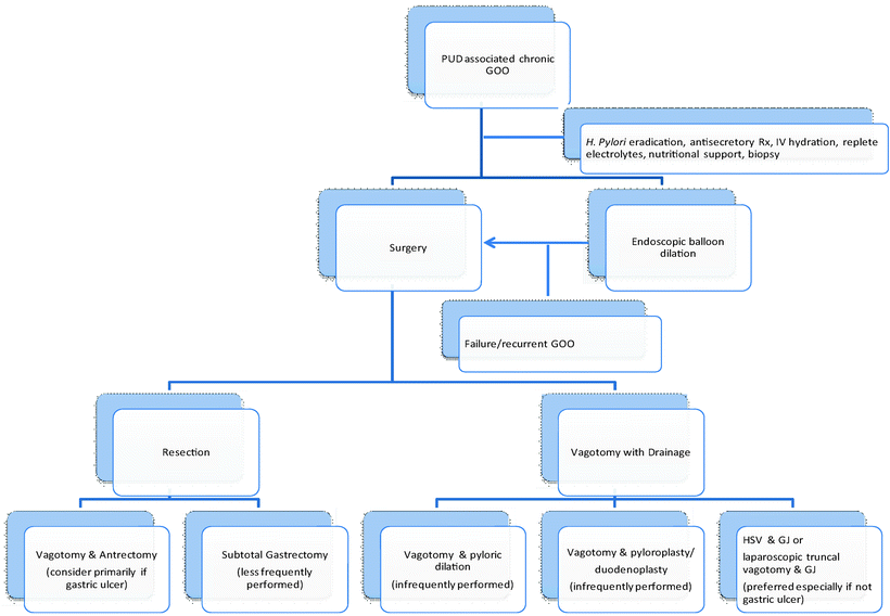
GOO from PUD is usually secondary to a combination of edema, spasm, fibrotic stenosis, and gastric atony [3]. While acute GOO secondary to edema or spasm will usually resolve within 48–72 h, with the ability to resume a regular diet within 96 h, chronic GOO from PUD is unlikely to respond to nonoperative measures [9, 10]. Management of GOO secondary to PUD can be divided into three categories that are not mutually exclusive: medical, endoscopic/fluoroscopic, and surgical (Fig. 18.2).


Fig. 18.2
Management of chronic PUD-associated GOO
Medical (Conservative) Management
Medical management includes nil per os (NPO) status, NG tube decompression, intravenous fluid rehydration, and antisecretory therapy via proton pump inhibitors (PPI) or histamine receptor type 2 (H2) blockers infused intermittently or continuously [10, 13]. Normal saline is the preferred initial crystalloid for resuscitation [9, 10]. Lost potassium needs to be replaced. To this end, serial monitoring of electrolytes and acid–base status is advisable. Serum gastrin levels may be obtained if concern for gastrinoma is present; however, use of antisecretory agents may interfere with test results [9]. Parenteral nutrition may also be indicated after initial resuscitation [10]. Nonsteroidal anti-inflammatory drugs (NSAIDs) should be stopped. Once initial resuscitation is completed, consideration should be given to performing esophagogastroduodenoscopy (EGD). A complementary UGI study or CT scan may also be performed once resuscitation is well under way [10].
The role of H. pylori in GOO is unclear. Reported rates of H. pylori positivity are between 33 and 47% in small studies [4, 21]. Nevertheless, testing for H. pylori should be performed, as its presence may predict successful balloon dilation decreased ulcer complication rate [21]. It has been noted that patients without H. pylori infection have a more severe ulcer diathesis. [21] It is hypothesized that patients without H. pylori infection may have chronic scarring that is less amenable to balloon dilation [21]. H. pylori infection can be diagnosed with a rapid urease breath test or on pathologic examination of samples [21]. While readily available, stool antigen testing and serologic testing are considered less accurate indicators of active infection [30]. If testing is positive, then eradication therapy should be commenced; this can be intravenous until enteral access is achieved. A variety of treatment regimens for H. pylori are available, including omeprazole, amoxicillin, and clarithromycin [21]. Of note, there are several case reports demonstrating resolution of GOO with H. pylori treatment, without recurrent GOO [8, 31]. These studies concluded that GOO associated with PUD is primarily because of edema and spasm and not cicatrization of the pyloric canal, and hence recommend prolonging a trial of medical management to 2 weeks, before proceeding with interventional techniques [8 , 31].
Medical management alone is generally favored in patients with first episodes of acute obstruction. Edema and gastritis are seen on endoscopy. This generally resolves quickly [9]. However, follow-up endoscopy is recommended to confirm H. pylori eradication and resolution of obstruction [9]. GOO unresponsive to medical therapy continues to be a problem in a small percentage of patients with PUD and is the main indication for surgery in a small percentage of patients requiring surgery for PUD [32]. Failure of medical management can be addressed via endoscopic/fluoroscopic balloon dilatation or surgery. The exact role of endoscopic balloon dilatation is still being defined. Surgery is recommended if there is concern for malignancy.
Endoscopic Management: Balloon Dilation
The primary therapeutic endoscopic modality for PUD-associated GOO is endoscopic balloon dilation (Table 18.1). It was first described by Benjamin et al., who used the principles of balloon catheter dilation in angiography to successfully perform through the scope (TTS) balloon dilation of a stenotic pylorus in a patient with GOO and acute myocardial infarction. Presently, balloon dilation is commonly done endoscopically; however the addition of fluoroscopy, with the use of contrast medium for balloon inflation, facilitates the procedure and may make it safer [21, 33]. Balloon dilation can also be performed under fluoroscopic guidance alone.
Table 18.1
Selected studies comparing stenting and GJ in malignant GOO
Ref. number | Year | n | Type | Results |
|---|---|---|---|---|
[103] | 2004 | 18 | Prospective, randomized Covered stent vs. open GJ | No statistically significant differences between the two groups in terms of morbidity, mortality, delayed gastric emptying, and clinical outcomes at 3-month follow-up |
[104] | 2004 | 36 | Prospective SEMS vs. open GJ | 100% of the patients that were alive in the stenting group could eat at 1 month 81% of the patients that were alive in the surgical group could eat at 1 month Shorter mean postoperative stay with stenting at 7.3 vs. 14.7 days Significantly less initial hospitalization cost with stenting, but the cost over the remaining lifetime was not significantly different between the stenting and surgery groups Advantage of stents in that they can be performed under conscious sedation |
[70] | 2005 | 47 | Retrospective Stent vs. open GJ | Comparable technical success rates but lower clinical success rates in the surgery group Lower morbidity and 30-day mortality rate in the endoscopic group |
[83] | 2005 | 22 | Retrospective review Stent vs. open GJ | 100% technical success rate in both groups 77.3% clinical success rate in both groups Major reasons for clinical failure included peritoneal dissemination, dysmotility, anastomotic dysfunction in GJ patients 75% of stent patients and 72.2% of GJ patients became independent of parenteral support No significant difference in the incidence of post-op complications Chemotherapy following stent insertion did not increase the risk of complications |
[105] | 2006 | 41 | Nonrandomized controlled Stent vs. open GJ | Stented patients achieved a significantly faster oral intake at an average of 2.4 days vs. 5 days for the surgical group Stenting group significantly shorter hospital length of stay at an average of 7.1 days vs. 11.5 days for the open group Significantly lower 30-day mortality rate of 16.6% in the stent group vs. 29.4% in the surgical group 68% of patients developed biliary obstruction |
[82] | 2008 | 50 | Prospective, observational | Median overall survival 64 days, did not differ significantly between patients treated with stents, GJ, or PEG/PEJ Significantly shorter median hospital length of stay with stenting at 2.5 days than other therapies Similar re-intervention rates between surgical GJ and stented groups at 3 months Acceptable QOL scores in both surgical GJ and stent groups |
[106] | 2010 | 39 | Multicentered, randomized Stent vs. open or laparoscopic GJ | No significant difference in survival between stent and surgery Hospital stay was significantly shorter in the stenting group at 7 days vs. 15 days in the surgical group Significantly shorter time to tolerance of oral intake in the stenting group, with a median of 5 days vs. 8 days By 2 months, food intake as measured by the GOOSS score was significantly better in the surgical group When including stent obstruction, there were significantly more major complications following stent placement Significantly more patients with stents required re-intervention for obstructive symptoms Hospital costs were significantly higher in the surgical group, primarily secondary to the longer hospital stay No significant differences in the health-related quality of life in follow-up between stenting and surgery |
Several factors need to be considered prior to embarking on balloon dilation. First, malignancy needs to be excluded. This may be done via a combination of endoscopy with biopsy and CT scan. When endoscopy is done for PUD-associated GOO, multiple and perhaps repeat or jumbo biopsies should be taken to exclude malignancy [28, 29]. Second, in patients with H. pylori infection, both eradication therapy and balloon dilation appear necessary to decrease the risk of recurrent GOO [4]. Third, long-term antisecretory/antacid therapy will also be needed to decrease the recurrence rate [4, 5, 20]. Fourth, while generally effective initially, endoscopic balloon dilation has a high recurrence rate [10, 11]. Greater than 80% of patients treated with balloon dilation will eventually require surgical intervention [10].
Once the decision has been made to perform balloon dilation, the size of the balloon must be carefully considered, as smaller balloons are associated with higher recurrence rates, whereas larger balloons (greater than 15 mm diameter) are associated with an increased perforation risk [21, 33, 34]. In deciding upon balloon size, it should be recalled that the normal adult pyloric canal diameter is about 15 mm [35].
Endoscopic Balloon Dilation Results, Complications, and Follow-Up
Successful dilation has been defined as an expansion of the obstructed segment to 10–15 mm diameter and resolution of symptoms [21, 36]. Failed balloon dilation may be secondary to long, tortuous strictures and severe fibrosis with anatomic distortion [5]. Following endoscopic dilation, the diet is advanced as tolerated. Overall, endoscopic balloon dilation has a very favorable safety profile. However, post procedure, patients should be monitored for bleeding or perforation [28]. Perforation may occur in up to 6% of patients [11]. If perforation is suspected, diagnostic choices are an upper GI study with water-soluble contrast or CT scan with oral contrast [28]. To decrease perforation risk, some authors have suggested graded dilations at 1–2-week intervals as needed [21, 28]. A third complication is pain. It is not uncommon, but is generally self-limited.
In the majority of patients, symptoms tend to improve rapidly following balloon dilation and patients are able to resume oral intake shortly thereafter [19–21, 36]. Further evidence of the efficacy of balloon dilation is provided by scintigraphic scanning, which has demonstrated improved gastric emptying [36]. However, the long-term results tend to be less favorable [10]. Several small studies (40 patients or less) reported outcomes that varied from a 36% recurrence rate at 2 years, to 70% recurrence rate at 3.5 years, to an 84% recurrence at a median follow-up of a little less than 4 years [19–21]. In a study with 40 patients, 30% were relieved with a single dilation, and 30% were referred for surgery [34]. Another small study found that all patients were asymptomatic at a median follow-up of 43 months using a combination of antisecretory therapy, endoscopic balloon dilation, and removal of etiologic factors; however, 91% of patients required a median of two dilations [19].
Given the high likelihood of recurrent ulceration/GOO and concern for underlying malignancy, patients undergoing endoscopic dilation should have long-term follow-up. Also, in H. pylori-positive patients, H. pylori eradication should be confirmed. Patients with recurrent or intractable symptoms of GOO despite multiple attempts at endoscopic therapy should be considered for surgical intervention [28]. Underlying malignancy should be considered in patients who develop rapid restenosis after dilation [5]. Factors predictive of referral for surgery include younger age, technical failure, need for multiple dilations, need for endoscopic intervention after 1 year, and a long duration of treatment course [10, 34]. Given these considerations, some authors feel that balloon dilation should be reserved as a temporizing measure or used in patients who are otherwise too ill to undergo surgical intervention [10, 30].
Surgery
Given the remarkable efficacy of PPI, H. pylori eradication therapy, and NSAID avoidance, the role of a major surgical resection and/or vagal resection for complicated PUD has become less clear [17]. While this may be in part because endoscopic and interventional radiologic techniques are comparatively recent inventions, the need for salvage surgical intervention if other therapies fail or to treat complications of other interventions cannot be argued [4, 37]. Surgery appears to be the gold standard against which all others are judged. For patients with recurrent symptoms after two balloon dilations, and/or those who are H. pylori negative, surgical evaluation is suggested [4]. The goals of surgery are to eliminate long-term antiulcer medication use and cure the GOO with a single procedure [5]. Given gastric dysmotility with chronic GOO, consideration can be given to placement of a jejunal feeding tube and decompressive gastrostomy [38].
Preoperative Care
Surgery is usually delayed for 5–7 days while the patient is rehydrated and electrolyte imbalances are resolved [22]. Stomach lavage/decompression for at least the day prior to surgery is advisable [22]. NGT decompression is continued intra- and postoperatively. Deep venous thrombosis (DVT) prophylaxis and perioperative antibiotics are administered according to Surgical Care Improvement Project (SCIP) guidelines.
Resective Surgery
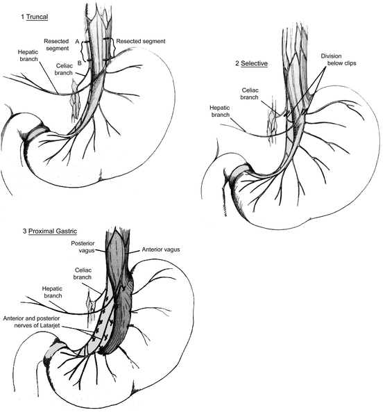
In selecting the operative intervention, factors that need to be considered are procedural morbidity, mortality, and ulcer/GOO recurrence rate. Some authors favor resective procedures, especially for gastric ulcer-associated GOO, while others do not [10]. Choices of surgical resection are procedural PUD-associated GOO include subtotal gastrectomy and antrectomy with vagotomy. Vagotomy is not necessary with subtotal gastrectomy. With antrectomy (also known as distal gastrectomy), whereby the distal 1/3 to 1/2 of the stomach are removed, a vagotomy is performed to further reduce acid secretion. Vagotomy may be truncal (at the esophageal hiatus, proximal to the hepatic and celiac branches of the vagus nerves) or selective (distal to the hepatic and celiac branches of the vagus nerves) (Fig. 18.3). Both subtotal gastrectomy and vagotomy with antrectomy (V&A) have higher perioperative morbidity rates but lower incidence of ulcer recurrence, as compared to non-resective procedures [39]. Specifically, V&A has a PUD recurrence rate of up to 1.5%, a 2% mortality, and significant morbidity [5]. The morbidity includes dumping syndrome in 25% of patients, although generally not severe and often resolving over time; alkaline reflux gastritis which affects 3–4% of patients and is persistent; diarrhea in up to 23% of patients; and nausea and bilious vomiting [5].


Fig. 18.3
Types of vagotomy. Figure reproduced by permission from Skandalakis L, Gray S, Skandalakis J. The history and surgical anatomy of the vagus nerve. Surgery, Gynecology & Obstetrics Journal, 1986;162:83 (ref. [108])
Following gastric resection, options for reconstruction include a Billroth I gastroduodenostomy, Billroth II gastrojejunostomy, or Roux-en-Y gastrojejunostomy. Data supporting Roux-en-Y reconstruction was provided by Csendes et al. in 2009 in a randomized study comparing Billroth II and Roux-en-Y anastomosis after partial gastrectomy with vagotomy [40]. They found significantly less symptoms, higher percentage of Visick I score (i.e asymptomatic) significantly more frequent normal distal esophageal endoscopic findings, significantly less frequent short segment Barrett’s esophagitis, and significantly more frequent normal gastric endoscopic findings in patients undergoing Roux-en-Y reconstruction as opposed to Billroth II reconstruction [40].
Vagotomy and antrectomy are often reported as preferred management options for GOO secondary to gastric ulcer [10]. However, it is important to note that in some complicated cases of GOO, significant scarring between the stomach, pancreas, and duodenum can make performing a resection difficult. Resection may leave a difficult duodenal stump. It also carries much higher morbidity and mortality rates.
Non-resective Surgery (Vagotomy with Drainage)
If non-resective surgery is decided upon for duodenal or gastric ulcer, then biopsies, especially in the case of gastric ulcer, should be performed to exclude malignancy [10]. Therapeutic alternatives include vagotomy (truncal or highly selective vagotomy [HSV]) with drainage (pyloric dilation, pyloroplasty, duodenoplasty, or gastroenterostomy). With HSV (also known as proximal gastric vagotomy or parietal cell vagotomy), the nerves of Latarjet (gastric divisions of the anterior and posterior vagus nerves) and Crow’s feet innervation to the antropyloric area are preserved (Fig. 18.3). As classically described for PUD management, an HSV is not performed with a drainage procedure because innervation to the pylorus is preserved; however, in the setting of GOO, HSV must be performed with a drainage procedure [41]. There is little data available on selective vagotomy with drainage for GOO.
One drainage procedure is pyloric dilation. In performing pyloric dilation, Hegar or Bakes’ dilators, a finger, or both can be used [5, 8]. A balloon-tipped catheter may also be used, especially if performing the procedure laparoscopically [38]. In a study of 30 patients with symptomatic PUD-associated GOO undergoing HSV with digital duodenal dilatation 90% of patients with initially symptomatic stenosis had no further problems with duodenal ulceration, with a follow-up time of up to 10 years [23]. Surgical dilatation carries a perforation rate from 0% to 27% [5]. Dumping and diarrhea have been found in up to 7% of patients, and delayed gastric emptying has been reported in 3–47% of patients [5]. Restenosis rates of up to 16% have been reported in the literature [5]. It should be noted that this may also be performed in the duodenum. Because of the availability of endoscopic balloon dilation, antisecretory therapy, and H. pylori treatment, this approach is currently used infrequently [10].
A second option advocated by some for drainage following vagotomy is pyloroplasty, where choices include Heineke–Mikulicz, Finney, or Jaboulay pyloroplasty [5]. For a Heineke–Mikulicz pyloroplasty, the pylorus is longitudinally incised and then closed transversely. Alternatively, depending on stricture location, a duodenoplasty may be indicated. In this case, a longitudinal incision of the stricture is followed by subsequent transverse closure [5]. Some authors argue against pyloroplasty as a drainage procedure as dissection of the obstructing segment or pylorus and closure of the duodenal stump can be challenging [10, 42]. Because of these reasons, and the availability of other techniques, vagotomy with pyloroplasty has fallen out of favor.
A third choice for drainage following vagotomy is gastroenteric anastomosis. The choice between pyloroplasty and gastrojejunostomy (GJ) is made in part by the appearance of the pyloric-duodenal area. A GJ is currently the favored approach. When a gastrojejunal anastomosis is chosen, decisions must be made as to whether the anastomosis will be retrocolic or antecolic, located on the posterior or anterior aspect of the stomach, and isoperistaltic or antiperistaltic. A retrocolic anastomosis will require the creation of a window in the gastrocolic omentum. In selecting the location of the gastroenteric anastomosis for benign disease, it is believed that an anastomosis with the posterior gastric wall facilitates drainage [43]. Furthermore, the anastomosis should be located on the most dependant part of the greater curve or antrum, as close to the pylorus as possible [22].
HSV with GJ is generally the preferred treatment for chronic GOO [13]. HSV maintains antral propulsive activity [41]. In comparing HSV with truncal V&A, complications such as delayed gastric emptying, gastric atony, dumping, alkaline reflux gastritis, diarrhea, cholelithiasis, and weight loss are less common with HSV because pyloric innervation is preserved [5, 10]. However, ulcer recurrence rates of 3–30% with HSV, depending on surgeon experience, are much higher than V&A, especially after greater than 10-year follow-up [5]. HSV is favored over V&A in poor-risk patients and/or those with problematic duodenums [10].
Two notable older trials compared open surgical management strategies in GOO. It should be noted that both trials included patients from the 1970s, around the time that H2 blocker use was in its early stages and well before PPI were widely marketed. In 1993, Csendes et al. published the results of a prospective randomized study comparing open HSV with GJ, HSV with Jaboulay gastroduodenostomy, and selective vagotomy (SV) with antrectomy in 90 patients with GOO secondary to duodenal ulcer [41]. As compared to HSV with Jaboulay gastroduodenostomy, on late follow-up, there were a significantly higher number of patients with HSV with GJ with Visick I score. They concluded that HSV with gastrojejunostomy as compared to the other two operations was the procedure of choice in GOO secondary to duodenal ulcer. Meanwhile Makela et al. analyzed 99 patients with GOO secondary to PUD and noted a 5% post-operative mortality rate [3]. They found an 11% restenosis rate in patients undergoing Billroth I reconstruction, 0% incidence in Billroth II reconstruction, and 4% after Roux-en-Y reconstruction. They also noted a 5% incidence of restenosis in those undergoing selective vagotomy and antrectomy. Finally, they found a 42% rate of restenosis amongst those undergoing HSV with pyloroduodenal dilatation, and consequently, they argued against its use.
Laparoscopic/Laparoscopically Assisted Surgery for GOO
While both laparoscopic and open surgical intervention is used, the trend has been towards a greater role for laparoscopic surgery in the management of GOO. Often cited reasons for favoring a laparoscopic over an open approach include less pain, less immobility, shorter hospital length of stay, smaller wounds, and a quicker return to activities of daily living [44]. While laparoscopic HSV with gastrojejunostomy may also be considered, the literature primarily promulgates truncal vagotomy with gastroenterostomy. Hence, this technique will be discussed. A retrospective study of 21 GOO patients comparing laparoscopic with open truncal vagotomy and gastrojejunostomy noted that laparoscopic surgery was associated with significantly reduced operating time, intraoperative blood loss, time to flatus, time to tolerance of semisolid diet, length of hospital stay [42]. While laparoscopic equipment costs more, these costs are generally believed to be offset by the shorter hospital stay [44]. In considering laparoscopic procedures, while intervention may be done purely laparoscopically, there are also a variety of laparoscopically assisted techniques which may offer some element of patient safety, especially for those who are not expert laparoscopic surgeons. In this regard, a 2005 study of 18 patients with GOO who underwent laparoscopic truncal vagotomy followed by an extracorporeal antecolic posterior gastrojejunostomy reported no mortality, no conversions, and a median hospital stay of 6 days [45]. This study noted a 16% incidence of postvagotomy diarrhea, which is slightly higher than that commonly reported. Similarly, extracorporeal antecolic anastomosis was performed in a study of 18 patients undergoing laparoscopic-assisted gastrojejunostomy with truncal vagotomy for cicatrizing duodenal ulcer with GOO [22]. This study also noted that no patient required conversion to a fully open procedure, no mortality, but one patient had an anastomotic leak requiring laparotomy. With a mean follow-up of 22.8 months, none of the patients developed recurrent obstruction.
Open Highly Selective Vagotomy with Gastrojejunostomy Technique: General Principles
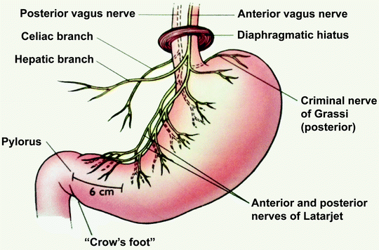
The general principles of HSV, as described in a standard surgical text, is briefly discussed [46]. The dissection is started at a point approximately 7 cm proximal to the pylorus, on the lesser curvature. The vagal nerve fibers distal to this point are preserved. The nerves of Latarjet are identified and encircled with vessel loops. The nerves emanating from the nerves of Latarjet along the lesser curvature along with associated blood vessels are ligated and divided. Dissection proceeds proximally to the esophagus, first dividing the anterior layer of nerves, followed by an irregular intermediate layer of nerves, and then the posterior layer of nerves. Finally, the esophagogastric junction is mobilized and branches to the stomach including the criminal nerve of Grassi, a branch of the posterior vagus nerve that supplies the cardia and can originate high in the mediastinum, are divided (Fig. 18.4). This generally involves clearing the distal 5 cm of the esophagus of vagal nerve fibers. A gastrojejunostomy is then performed near the antrum to the posterior wall of the stomach, along the greater curvature of the stomach using a gentle loop of jejunum [41].


Fig. 18.4
Criminal nerve of Grassi. Reproduced by permission from Diesen D, Haney J, Pappas J. Laparoscopic management of peptic ulcer disease. In: Pappas T, Pryor A, Harnisch M, eds. Atlas of laparoscopic surgery. 3rd ed. New York: Current Medicine; 2008:65–79 (ref. [109])
Laparoscopic/Laparoscopic-Assisted Truncal Vagotomy with Gastrojejunostomy Technique
The technique, as described by several authors, follows [39, 43, 45]. The patient is placed in reverse Trendelenburg, in the Lloyd-Davis position, with the operating surgeon standing between the patient’s legs. The peritoneal cavity is insufflated with carbon dioxide and several additional ports are inserted. The left lobe of the liver is retracted medially; subsequently the esophagus is identified by palpation of the previously placed nasogastric tube. The phrenosesophageal ligament is divided. The anterior vagus nerve is identified, clipped proximally and distally, transected, and the intervening segment is submitted for frozen section pathologic confirmation. Attention is then directed to the posterior vagus nerve; dissection in the peri-esophageal region between the right crus and esophagus facilitates exposure; again the main trunk is clipped and divided, and the intervening segment is submitted for frozen section pathologic analysis [39]. It is important not to carry the dissection too far towards the liver or too low over the gastric fundus [43]. A search is then made for additional branches of the vagus nerve, including the criminal nerve of Grassi [39, 43]. At least 5 cm of the distal esophagus is cleared of vagal fibers [39]. While the principles of laparoscopic vagal resection are the same as those in open surgery, with its magnified view, caution must be exercised to ensure that longitudinal esophageal muscles are not mistaken for the vagal nerves [43]. Gastrojejunostomy is performed using a loop of the jejunum that gives the shortest afferent loop, generally 30–40 cm from the ligament of Treitz [39, 43]. Ultrasonic dissection may be used to create the gastrostomy and jejunostomy [39]. The gastroenteric anastomosis can be completed intracorporeally or extracorporeally, using a gastrointestinal anastomosis stapling device or suture (Fig. 18.5). The opening at the anastomosis is then closed with suture or a stapler. All fascial incisions greater than 5 mm in diameter should be closed, unless a radially dilating type port is used. A radially dilating port may allow a slightly larger fascial incision to be left open.


Fig. 18.5
Laparoscopic GJ diagram. (a) After placement of stay sutures, an opening is made in both the stomach and jejunum. (b) An endoscopic stapler is introduced into the adjacent stomach and jejunum. (c) The open end of the anastomosis is closed with sutures. Figure reproduced permission from Brune IB, Feussner H, Neuhaus H, Classen M, Siewert JR. Laparoscopic gastrojejunostomy and endoscopic biliary stent placement for palliation of incurable gastric outlet obstruction with cholestasis. Surg Endosc. 1997;11(8):834–837 (ref. [110])
Operative Pearls
In operating on the obstructed stomach, every effort should be made to evacuate fluid and gases from the stomach prior to entering the gastrointestinal tract [47]. Caution must be exercised in the use of electrocautery, as explosions have been reported secondary to retained gases [47]. When performing operations on the stomach for GOO, consideration should be given to performing hand-sewn anastomoses or using a greater tissue depth staple cartridge, to ensure adequate purchase on the hypertrophied stomach [10].
The appearance of the pylorus and proximal duodenum influences surgical intervention. Extensive inflammation may make a Billroth I procedure impossible; it may also yield a difficult duodenal stump with a Billroth II reconstruction [17, 30, 42]. In those patients with a difficult duodenal closure, consideration can be given to performing a tube duodenostomy and perihepatic drainage [4]. However, it is better to avoid this situation altogether and perform a vagotomy with gastroenterostomy [4].
Postoperative Care, Complications, and Follow-Up After Surgery
Surgery is highly effective in the management of GOO. Postoperatively, the patient is admitted to a monitored floor. Fluid and electrolyte status need to be followed closely. Nutritional support should be continued. While the duration of NGT decompression is controversial, it should be recalled that patients with GOO may need NGT for a longer period of time secondary to delayed gastric emptying from chronic gastric atony as well as vagotomy. Follow-up with the surgeon is generally performed by about 2 weeks post discharge. A second follow-up visit may occur several weeks later, and then as needed to address any complications that may arise. Long-term follow-up is usually with the primary care physician and/or gastroenterologist.
Complications that may occur postoperatively include atelectasis, fever, arrhythmia, myocardial infarction, DVT, pulmonary embolism (PE), pneumonia, bleeding, wound infection, and in the case of an anastomosis, an anastomotic leak. Long-term complications of truncal vagotomy include gastric stasis, bilious vomiting, diarrhea, and dumping [42]. The literature suggests a 5–16% incidence of diarrhea following vagotomy [45]. Diarrhea from the dumping syndrome is secondary to massive outpouring of fluid from the vascular compartment into the bowel lumen, because early gastric emptying produces hyperosmolar intraluminal contents [45]. Additional complications of commonly performed PUD operations included anastomotic obstruction/stenosis, obstruction of the afferent or efferent loops, delayed gastric emptying, and marginal ulceration.
PUD Conclusions
In conclusion, while PUD-associated acute GOO often resolves with conservative measures, chronic GOO will generally require intervention. While the role of H. pylori in GOO is unclear, infection must be assessed and eradication therapy administered if it is found. Antisecretory medications are administered and malignancy must be excluded. Depending on patient preference and in poor-risk surgical candidates, it appears reasonable to offer a trial of endoscopic balloon dilation as it provides a rapid relief of symptoms and has a favorable safety profile, with the understanding that there is a high long-term recurrent GOO risk and long-term antisecretory therapy will be needed. In good-risk patients who are H. pylori negative, fail endoscopic treatment, wish to avoid long-term antisecretory medication treatment, or desire definitive treatment, surgical intervention should be performed. In deciding amongst the myriad of operations, one algorithm to consider is to perform vagotomy and antrectomy in the patient with a gastric ulcer with good performance status and simple anatomy. If the gastric ulcer patient has a limited performance status, then vagotomy with gastroenterostomy should be performed. If the patient has a duodenal ulcer, consider performing HSV with drainage or truncal vagotomy with drainage. While either technique can be performed laparoscopically, there is presently very little literature available on laparoscopic HSV with drainage for GOO. The decision between the two would depend on the surgeon’s familiarity with the procedures; it has been reported that surgeons today may have limited operative experience with GOO [48]. Additional factors to guide decision-making are informed consent from the patient, regarding side effects from surgery and risk of recurrent ulceration. As previously discussed, HSV with drainage has fewer side effects but a higher risk of recurrent PUD than truncal vagotomy with drainage.
Other Benign Causes of Goo
Inflammatory Conditions
Several local and systemic inflammatory conditions can also cause GOO. Local conditions include acute and chronic pancreatitis, pancreatic pseudocyst, and obstruction secondary to cholecystoduodenal fistula with calculous cholecystitis (Bouveret’s syndrome) [9, 49, 50]. Systemic inflammatory conditions include Crohn’s disease. While gastroduodenal involvement in Crohn’s disease is rare, with an incidence of about 5%, stenosis was noted in 78% of 54 patients in a small series of patients with gastroduodenal disease [51]. Other conditions include Behcet’s disease and systemic lupus erythematosus [9]. Finally, eosinophilic gastroenteritis can result in intestinal obstruction due to muscular infiltration with eosinophils [15].
Stay updated, free articles. Join our Telegram channel

Full access? Get Clinical Tree


