Laboratory evaluation: Laboratory signs of dehydration may include a rising hematocrit (acute blood loss will not result in an acute change of hematocrit), metabolic acidosis, a urinary specific gravity greater that 1.010, a urinary sodium less than 10 mEq/L, a urinary osmolality greater than 450 mOsm/kg, hypernatremia, and a ratio of blood urea nitrogen to creatinine greater than 10:1.
Hemodynamic measurements: Central venous pressure (CVP) measurements alone do not provide accurate assessment of volume status. Pulmonary artery (PA) catheter placement can obtain CVP, right ventricular pressure, and PA and PA occlusion pressures, but the accuracy of these figures in relation to intravascular volume status can be difficult to interpret and relies on multiple factors, including normal pulmonary physiology, intact cardiac valve function, normal intrathoracic pressures, and so on. Other options include arterial pulse contour analysis or transesophageal echocardiography (TEE). TEE may give the best estimate of volume status because it offers direct visualization of ventricular filling and avoids complications associated with PA catheter use. In short, all hemodynamic measurements need to be interpreted in the context of the clinical setting.
Intravenous Fluids
Crystalloid solutions: Largely considered the initial resuscitation fluid in patients with hemorrhagic and septic shock, burns, and traumatic brain injuries. A general rule is to replace what is being lost. In other words, for losses involving water (“nothing by mouth” patients on the floor), replacement with hypotonic solutions is appropriate for maintenance. If losses involve water, electrolytes (i.e., blood loss), replacement is with isotonic electrolyte solutions. Glucose is rarely added in the intraoperative setting, although pediatric patients are prone to hypoglycemia and often need a glucose source with their fluids. Normal saline, when given in large volumes, can produce a dilutional hyperchloremic acidosis.
Colloid solutions: The intravascular half-life is between 3 and 6 hours. These solutions are derived either from plasma proteins or synthetic glucose polymers. Crystalloid versus colloid resuscitation continues to be an ongoing debate, but the use of albumin (5% and 25%) is justified in the presence of hypoalbuminemia or large burns (large protein loss). The synthetic colloids run the risk of antiplatelet effects and should not be administered over 20 mL/kg/day. The dextrans have also been found to be antigenic and can produce anaphylactoid reactions.
Perioperative Fluid Therapy
Normal Maintenance
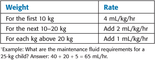
Estimating Maintenance Fluid Requirements1

Preexisting Deficits
• Calculation: The total fluid deficit can be derived from multiplying the maintenance rate by the length of the fast. Abnormal fluid losses frequently contribute to preoperative deficits. Ideally, all deficits should be replaced preoperatively.
• ABO system: Determined by the presence or absence of A or B surface antigens. (Type A has A surface antigen, and type O has neither A nor B surface antigen.) Within the first year of life, antibodies are produced against the missing antigens (anti-B antibody for the type A blood group).
• Rh system: There are approximately 46 rhesus group surface antigens. Patients with the D rhesus antigen are considered Rh positive. An Rh-negative patient will only develop antibodies against the D antigen after exposure to Rh-positive blood (i.e., transfusion, pregnancy).
• Other red blood cell (RBC) antigen systems: Include Lewis, Duffy, Kell, Kidd, and so on. Alloantibodies against these antigens rarely cause serious hemolytic reactions but can be tested with a crossmatch.
• ABO-Rh testing (“type”): Recipient RBCs are tested with a serum known to have antibodies against A and B to determine the patient’s blood type. Then the patient’s serum (plasma) is mixed with RBCs with a known antigen type to confirm the type. Anti-D antibodies are also added to determine Rh status. If the patient is Rh negative, the plasma is mixed against Rh-positive RBCs to determine if the patient has anti-D antibodies (particularly important in pregnancy).
• Antibody screen (“screen”): Tests the serum of the patient or donated blood to determine if any clinically significant antibodies (those that are associated with non-ABO hemolytic reactions) exist. An indirect Coombs test is carried out, which involves mixing the patient or donor serum with a panel of RBCs with known antigenic composition. If specific antibodies are present, they will attach to the antigen and ultimately agglutinate that sample.
• Crossmatch (“cross”): Mimics what would happen in the actual transfusion. The donor RBCs are mixed with recipient serum to look for any untoward reaction. This step is rarely done in most medical centers because a “type and screen” reduces the likelihood of a hemolytic reaction to one in 10,000. If the patient develops a positive antibody screen result, a crossmatch will be performed before transfusion.
• Emergency transfusions: If recipient type and Rh status are not known, type O Rh-negative (universal donor) blood may be used.
Stay updated, free articles. Join our Telegram channel

Full access? Get Clinical Tree


