Chapter 22
Extremity Compartment Syndromes
Col. (retd.) Mark W. Bowyer
Chapter Overview
All clinicians caring for critically ill patients must be able to recognize and treat (or refer for treatment) Compartment Syndrome (CS) of the extremities. CS results from a variety of etiologies (traumatic and non-traumatic) with the final common pathway being increased compartmental pressure that exceeds the arterial inflow with resultant ischemia and necrosis. Failure to identify and treat CS in a timely fashion is associated with preventable morbidity and mortality, and is a common source of litigation. The diagnosis of CS is largely clinical, but measurement of compartment pressures may be useful in patients with equivocal findings or altered level of consciousness. The below-knee lower extremity is most commonly affected, followed much less frequently, by the forearm, thigh, foot, and hand. This chapter will review the pathophysiology, epidemiology, diagnosis, relevant anatomy, and treatment of CS, emphasizing the proper performance of a fasciotomy, and the complications associated with this vital limb and potentially life-saving procedure.
Introduction
CS is a condition in which increased pressure within a limited space compromises the circulation and function of affected tissues.1,2 This limb and potentially life-threatening condition may result from several possible etiologies (Table 1),1,3,4,7 with which every clinician should be familiar. Failure to identify and treat CS properly, leads to tissue necrosis, permanent functional impairment, possible amputation, and potential renal failure and death.1,3,4 In a nine-year review of extremity trauma, Feliciano et al.5 found that 75% of amputations in the lower extremity were related to a delay in performing fasciotomy or an incomplete fasciotomy.
Table 1. Factors implicated with the development of acute limb CS.1,3,4,7
Restriction of compartment size | Increased compartment volume |
| From Hemorrhage |
| Fractures |
Casts | Vascular Injury |
Splints | Drugs: (anticoagulants) |
Burn Eschar | Hemophilia; Sickle Cell |
Tourniquets | From Muscle Edema/Swelling |
Tight Dressings | Crush — trauma, drugs or alcohol |
Fracture Reduction | Rhabdomyolysis/Blast injury |
Closure of Fascial Defects | Sepsis |
Incomplete Skin Release | Exercise induced |
Military Antishock Trousers | Envenomation or Bee Sting |
Prolonged Extrication Trapped Limb | Massive resuscitation |
Localized External Pressure | Intra-compartmental fluid infusion |
Long Leg Brace | Phlegmasia caerulea dolens |
Automated BP monitoring | Electrical burns |
Malpositioning on OR table | Reperfusion Injury |
| Post Partum Eclampsia |
Not only is disability resulting from CS of great consequence to the patient, but failure to diagnose or properly treat CS is one of the most common causes of medical litigation.6 Bhattacharyya and Vrahas6 reported an average indemnity payment of $426,000 in nine cases settled between 1980 and 2003 in Massachusetts, and awards as high as $14.9 million have been made in cases of missed CS.
Optimal outcomes result from early recognition of CS and aggressive, properly performed fasciotomy. Proper fasciotomy requires extensive knowledge of the anatomical landmarks and anatomy of the muscle compartments of the extremities.
Pathophysiology
Groups of muscles and their associated nerves and vessels are surrounded by thick fascial layers that define the various compartments of the extremities which are of relatively fixed volume. CS occurs either when compartment size is restricted or when compartment volume is increased. It is imperative that all clinicians working in critical care settings be aware of the numerous non-traumatic causes (Table 1) of extremity CS, especially sepsis, massive resuscitation, and reperfusion as the diagnosis of CS in these settings is often delayed, as it is frequently not considered by many otherwise well-trained physicians.
As pressure increases, venous flow decreases and narrows the arteriovenous perfusion gradient, resulting in diminished blood flow. This condition is self-perpetuating, leading to a continuous loop that must be broken with the timely initiation of definitive care. Cellular hypoxia is the final common pathway of all compartment syndromes. As ischemia continues, irreparable damage to tissue ensues and myoneural necrosis occurs. Development of CS depends on many factors, including the duration of the pressure elevation, the metabolic rate of the tissues, vascular tone, associated soft tissue damage, and local blood pressure.11 Nerves demonstrate functional abnormalities (paresthesias and hypoesthesia) within 30 minutes of ischemic onset. Irreversible functional loss will occur after 12 to 24 hours of total ischemia.1 Muscle shows functional changes after 2 to 4 hours of ischemia with irreversible loss of function beginning at 4 to 12 hours.1 Clinically, there is no precise pressure threshold and duration above which significant damage is irreversible and below which recovery is assured.
Tissue previously subjected to intervals of ischemia is especially sensitive to increased pressure. Bernot and colleagues8 showed that tissue compromised by ischemia prior to an elevated compartment pressure has a lower threshold for metabolic deterioration and irreversible damage. Polytrauma or otherwise critically ill patients with low blood pressures can sustain irreversible injury at lower compartment pressures than patients with normal blood pressures, and a very high index of suspicion should be maintained in this group.
Epidemiology/risk factors
Given the consequences of missing a CS, it is important to identify the population at risk. Trauma is the major cause of extremity CS requiring fasciotomy. In a 10-year retrospective review of over 10,000 trauma patients sustaining extremity injury, Branco et al., described a fasciotomy rate of 2.8%.3 During this period 315 fasciotomies were performed on 237 patients with 68.4% done below the knee, 14.4% on the forearm, and 8.9% on the thigh. In a review of 294 combat injured soldiers undergoing 494 fasciotomies, Ritenour et al. reported the calf as the most common site (51%) followed by the forearm (22.3%), thigh (8.3%), upper arm (7.3%), hand (5.7%), and the foot (4.8%).9
Branco et al.3 found that incidence of fasciotomy varied widely by mechanism of injury (0.9% after motor vehicle collision to 8.6% after a gunshot wound). Additionally, the need for fasciotomy was related to the type of injury ranging from 2.2% incidence for patients with closed fractures up to 41.8% in patients with combined venous and arterial injuries. Young males, with penetrating or multi-system trauma, requiring blood transfusion, with open fractures, elbow or knee dislocations, or vascular injury (arterial, venous, or combined) are at the highest risk of requiring a fasciotomy after extremity trauma.3
Diagnosis
Diagnosis depends on a high clinical suspicion and an understanding of risk factors, pathophysiology, and subtle physical findings. The diagnosis of CS is often based on subtle changes in symptoms and vague clinical findings. Time to diagnosis and treatment are the most important prognostic factors. Incomplete knowledge of the natural history and signs and symptoms primarily account for delays in diagnosis.10 The aim is to recognize and treat raised intra-compartmental pressure before irreversible cell damage occurs.
Numerous authors have stated that the diagnosis of CS is a clinical diagnosis.1,7,10,11 The classically described five “Ps” — pain, pallor, paresthesias, paralysis, and pulselessness are said to be pathognomonic of CS. However, these are usually late signs and extensive and irreversible damage may have taken place by the time they are manifested. In the earliest stages of CS, patients may report some tingling and an uncomfortable feeling in their extremity followed closely by pain with passive stretching of the muscles of the affected compartment. The most important symptom of CS is pain greater than expected due to the injury alone.
Nerve tissue is affected first by the subsequent hypoxia causing pain on passive motion seen early in the development of CS, sparing distal pulses until late in the course.7 The loss of pulse is a late finding, and the presence of pulses and normal capillary refill do not rule-out CS! The presence of open wounds does not exclude CS. In fact, the worst open fractures are actually more likely to have a CS.
All clinical signs have inherent drawbacks in making the diagnosis. Pain is an unreliable and variable predictor, and the pain of the obvious injury can mask that of an impending CS and cannot alone be relied upon for diagnosis. Wide consensus in the literature suggests that the clinical features of CS are more useful by their absence in excluding the diagnosis, than when they are present in confirming the diagnosis.
Since clinical findings may be absent in patients with altered sensorium (common in the intensive care setting), under the influence of drugs or alcohol, distracting injuries, or paralysis, many authors advise using tissue pressure measurements as an adjunct to clinical findings.12 There are also some who advocate the use of compartment pressure measurement as a principle criterion for the diagnosis of CS.
In actual practice, tissue pressure (compartment pressure) measurements have a limited role in making the diagnosis of CS. However, in polytrauma patients with associated head injury, drug and alcohol intoxication, intubation, spinal injuries, use of paralyzing drugs, extremes of age, unconsciousness, or low diastolic pressures, measuring compartment pressures may be of use in determining the need for fasciotomy.
The pressure threshold for making the diagnosis of CS is controversial. A number of authors recommend 30 mmHg, and others cite pressure as high as 45 mmHg. Many surgeons use the “Delta-P” system. The compartment pressure is subtracted from the patient’s diastolic blood pressure to obtain the Delta-P. Whitesides in 1975 proposed that muscle was at risk when the compartment pressure was within 10–30 mmHg of the diastolic pressure.12 If the Delta-P is less than 30, the surgeon should be concerned that a CS may be present. For instance, if the diastolic blood pressure was 60 and the measured compartment pressure was 42 (60 − 42 = 18), the “Delta-P” would be 18 and the patient is likely to have CS.
A variety of techniques have been used to measure compartment pressures but many suffer from the cumbersome nature of setting them up and user variability. A commercially available portable hand-held, self contained, electronic pressure monitor with a digital display is available (Stryker® Intra-Compartmental Pressure Monitor System, Stryker® Surgical, Kalamazoo, Michigan) has replaced most less reproducible devices as the current standard (Fig. 1). An alternative approach is to use an 18-gauge needle attached to a side-port arterial line set-up inserted into the compartment.
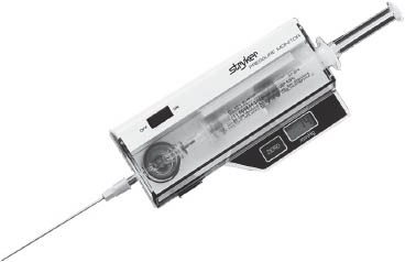
Fig. 1. Intra-Compartmental Pressure Monitor System manufactured by Stryker® Surgical, Kalamazoo, Michigan.
Source
Related posts:


Stay updated, free articles. Join our Telegram channel

Full access? Get Clinical Tree


