Basic Emergency Ultrasound
 Key Points
• Emergency ultrasound is frequently used by emergency medical providers for the management of patients in the emergency department for a variety of indications.
• Emergency ultrasound focuses on answering a specific question that has an impact on patient care.
• Applications for emergency ultrasound are expanding rapidly and having a positive impact on patient care.
Introduction
Distinction must be made between EUS and ultrasound performed by other specialties. The American College of Emergency Physicians (ACEP) has defined EUS as “the medical use of ultrasound for the bedside diagnostic evaluation of emergency medical conditions and diagnoses, resuscitation of the acutely ill, critically ill or injured, guidance of high risk or difficult procedures, monitoring of certain pathologic states and as an adjunct to therapy.”1 EUS is performed or directly supervised by emergency physicians in a variety of settings. Importantly, EUS is primarily distinguished from consultant-performed ultrasound in that it is a goal-directed, focused ultrasound examination.2 In short, EUS seeks to answer a brief or “yes or no” question. For example, in a patient with a complaint of abdominal pain and a positive urine pregnancy test, EUS seeks to answer the question “Is an intrauterine pregnancy present?” to guide diagnosis and treatment of the patient. Ultrasound performed by a consultant answers the question in a more expansive manner. For example, in the aforementioned patient, ultrasound performed by radiology or obstetrics and gynecology seeks to evaluate all the organs of the pelvis and to describe any abnormalities identified.
What We Are Looking For
The most common uses for EUS are listed in Box 1. Additionally, the ACEP released the Emergency Ultrasound Imaging Criteria Compendium in 2006,3 which further elucidates and defines the indications, technique, and interpretation of each of the eight primary types of bedside ultrasound. Although this list is extensive, applications for EUS continue to be refined and grow as emergency physicians respond to the challenges of patient care.
Physics of Ultrasound
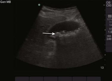
Tissues that are similar to water transmit sound waves very well and reflect less sound back to the transducer. Examples of these types of tissue in the body are the bladder, blood within the arteries and veins, and the vitreous humor of the eye. These tissues are seen as black or anechoic (without echoes) on the screen (Fig. 1). Conversely, tissue that has less water and is more dense reflects most or all of the sound waves that strike it. Examples of these types of tissue in the body are bones, tendons, and gallstones. These tissues are seen as white or hyperechoic (a lot of echoes) on the screen (Fig. 2). Continuing this logic, all other tissues that are somewhere in the middle of these textures fall into the range of shades of gray (Fig. 3). Objects that are mostly solid but contain some water are a lighter shade of gray, whereas objects that contain more water but are somewhat solid are a darker shade of gray.
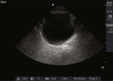
Fig. 1 Ultrasound image of the bladder.
Note that urine in the bladder, which is similar to fluid or water, is black or anechoic.