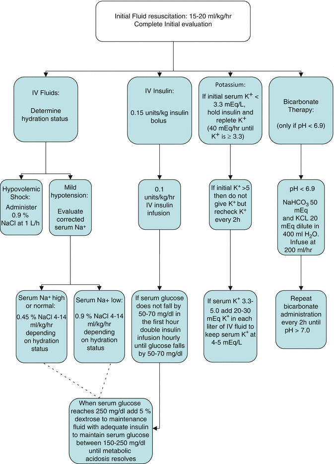
Fluid replacement therapy is usually initiated with isotonic saline. The initial infusion rate should be 15–20 ml/kg/h for the first 3–4 h (approximately 1.0 to 1.5 l/h for average sized adults), with a maximum of 50 ml/kg within the first 4 h.
After the initial fluid resuscitation, the choice of fluid replacement depends on the serum electrolytes, the serum glucose, and urine output. The sodium level should be adjusted for the serum glucose concentration [10].


If the corrected serum sodium level is normal or elevated, then 0.45 % NaCl should be used in place on 0.9 % NaCl to better correct the free water deficit. However, if the corrected serum sodium is low or the urine output is suboptimal, a persistent intravascular volume deficit is likely and 0.9 % NaCl should be continued. Once the serum glucose reaches 250 mg/dl, 5 % dextrose should be added to the IV fluids to maintain a serum glucose between 150–200 mg/dl until metabolic acidosis resolves.
Insulin
Insulin is typically administered as an IV bolus of 0.1-0.15 units/kg followed by an infusion of 0.1 units/kg/h. Prior to initiating insulin therapy, potassium must be repleted to at least 3.3 mEq/L, as insulin will shift potassium intracellularly, worsening hypokalemia and potentially causing cardiac arrhythmias. The target reduction in glucose levels is 50–70 mg/dL each hour. If the glucose does not fall by at least 50 mg/dL in the first hour, the insulin infusion should be doubled every hour until the desired rate of glucose decline is achieved. Insulin therapy should not be discontinued until metabolic acidosis resolves and the anion gap normalizes.
Electrolyte Repletion
If the initial serum potassium is 3.3–5.0 mEq/L, 20–30 mEq of potassium should be added to each liter of fluid. If the initial serum potassium is elevated, supplemental potassium should not be given and the serum potassium should be monitored every 2–3 h. The serum potassium should be maintained between 4–5 mEq/L.
Routine phosphate repletion is not recommended given the absence of clear benefit and its association with hypocalcemia and other electrolyte disturbances [11–13]. Most experts, however, recommend repletion when serum phosphate concentration falls below 1.0 mg/dl to prevent skeletal muscle weakness and respiratory failure.
Magnesium deficiency can result in cardiac dysrhythmias, seizures, muscle spasm, and paresthesia. Magnesium should be replaced when serum magnesium levels fall below 1.2 mg/dl.
Treatment of Precipitating Condition
While non-adherence to insulin therapy is often the precipitating factor in DKA, other causes such as infections and additional acute medical conditions (e.g., pancreatitis, myocardial infarction, etc.) should be actively considered [6, 14]. If a source of infection is suspected, concurrent treatment with appropriate antimicrobial therapy should not be delayed, especially if signs of shock are present.
Most patients with DKA will have a leukocytosis corresponding to the degree of acidosis; therefore the white blood cell count alone may not be a reliable predictor of infection. In one study of patients presenting to the emergency department with DKA, the presence of a significant number of immature band forms of leukocytes (i.e., bandemia) on the cell differential had a high sensitivity and specificity for the presence of serious infection [15]. Patients with significant bandemia should be considered to have a clinically relevant infection until proven otherwise.
Complications
The most feared complication of DKA is cerebral edema; however the vast majority of cases are seen in children. There have been case reports of adults developing cerebral edema after aggressive fluid resuscitation [16, 17]. Typical features include headache and lethargy followed by altered mental status, respiratory depression, seizures, and coma. Treatment is generally supportive. Mannitol and hypertonic saline have been used successfully to treat cerebral edema in DKA [18], although there are no controlled trials to support this practice.
Non-cardiogenic pulmonary edema is another potential complication of DKA treatment [19]. The pathophysiology may be related to decreased oncotic pressure and increased pulmonary capillary permeability [20–22]. Appropriate fluid resuscitation should not be withheld in patients with pulmonary edema as their intravascular volume may still be low (Fig. 47.1).
Evidence Contour
Bicarbonate Infusion
Use of bicarbonate in DKA remains controversial [22]. Prospective trials have not demonstrated any benefit or harm with bicarbonate therapy in patients with moderate metabolic acidosis (pH > 6.9) [23, 24]. Nonetheless, severe metabolic acidosis can impair cardiac contractility, reduce oxygen delivery by shifting the oxyhemoglobin dissociation curve, and cause organ dysfunction. Although there are no controlled trials supporting bicarbonate administration, most experts recommend initiating bicarbonate therapy when the pH is less than 6.9. Bicarbonate should be given as an infusion of 100 mmol sodium bicarbonate in 400 ml sterile water with 20 mEq of potassium over 2 h. The pH should be monitored and bicarbonate stopped once pH is greater than 7.0 [1].
Site of Care
Classically DKA has been treated with intravenous insulin, often in an intensive care unit setting. With current pressures to reduce costs and lengths of stay while maintaining optimal outcomes, much attention has been focused on alternative sites of care for DKA. Several studies have demonstrated the safety and feasibility of treating mild to moderate DKA with subcutaneous insulin analogues. One randomized, prospective trial demonstrated a significant reduction in hospital charges with the use of hourly subcutaneous insulin lispro administered on the general medical floor when compared to the use of an insulin infusion in the ICU, without an increase in adverse outcomes [25]. Other trials have also demonstrated the safety and efficacy of this approach [26].
Stay updated, free articles. Join our Telegram channel

Full access? Get Clinical Tree


