95 Cranial Nerve Disorders
• The 12 cranial nerves supply motor and sensory innervation to the head and neck.
• Cranial nerve disorders generally cause visual disturbances, facial weakness, or facial pain or paresthesias, depending on the nerve or nerves involved.
• Trigeminal neuralgia and Bell palsy are common cranial nerve disorders.
• A thorough history and physical examination should focus on assessing the potential for trauma (skull fracture), tumor, cerebrovascular accidents, vascular derangements (aneurysm, dissection, thrombosis), and infection (meningitis, abscess).
• The presence of concomitant focal neurologic or systemic signs should heighten suspicion for a central rather than a peripheral cause of the neurologic dysfunction.
Perspective
The 12 cranial nerves provide motor and sensory innervation to the head and neck. Some nerves serve purely motor functions (cranial nerves III, IV, VI, XI, and XII), some serve purely sensory functions (cranial nerves I, II, and VIII), and the remainder serve mixed motor and sensory functions (cranial nerves V, VII, IX, and X).
In addition to somatic and visceral sensory components, the cranial nerves provide the special sensory functions of sight, smell, hearing, taste, and balance. Understanding the functions of individual cranial nerves aids in recognition of patterns of the clinical syndromes classically associated with disorders of specific cranial nerves.
Epidemiology
Cranial neuropathies are a heterogeneous group of disorders with a variety of causes. Trauma is a common cause, and diabetes and hypertension are common comorbid conditions. Cranial nerves I, VI, and VII are the most frequently affected after minor head trauma.1 Trigeminal neuralgia is a common cause of facial pain that affects approximately 4.5 per 100,000 individuals; women are affected twice as often as men, and it is more common in those older than 60 years.2 Trigeminal neuralgia can be severely debilitating and has been termed the “suicide disease.”3 Bell palsy is the most common cause of acute facial paralysis worldwide. The peak age at incidence has been reported to be between 15 and 45 years,4 but other investigators have noted an increased incidence in individuals older than 70.5,6 Pregnant women and patients with diabetes have an associated increased incidence of the disease. A familial association of Bell palsy is noted in 4% of cases,4 and it can cause both significant psychologic and physical morbidity.
Pathophysiology
Cranial Nerve I (Olfactory Nerve)
Anatomy
Cranial nerve I is a special sensory nerve that provides the sense of smell. Inhaled scents are detected by the olfactory epithelium lining the nasal cavity and transmitted to the olfactory bulb, which lies adjacent to the cribriform plate of the ethmoid bone. Olfactory sensations are relayed from the olfactory bulb to the brain via the olfactory tract.
Presenting Signs and Symptoms
The patient should be questioned about a history of head trauma. An anteroposterior skull fracture parallel to the sagittal suture or an anteroposterior shearing injury can tear the olfactory fibers traversing the cribriform plate and lead to disruption of the synapses from the olfactory epithelium to the olfactory bulb.
A frontal lobe mass such as a tumor, meningioma, or abscess can compress the olfactory bulb as well, but the signs and symptoms associated with such masses tend to be more subacute.
Treatment
Treatment depends on the presence of concomitant injury. Basilar skull fracture and cerebrospinal fluid rhinorrhea associated with trauma require immediate neurosurgical consultation. A subacute mass or abscess should be managed in consultation with neurosurgery, depending on the acuity of the findings. Patients with anosmia secondary to trauma and normal findings on head computed tomography (CT) can referred to neurology or neurosurgery for outpatient follow-up.
Cranial Nerve II (Optic Nerve)
Anatomy
Visual stimuli are transmitted from the retina to the optic nerve through the optic chiasm to the lateral geniculate nucleus in the thalamus, where they synapse. From there, impulses are transmitted along the optic radiations (geniculocalcarine tracts, including the Meyer loop) to the primary visual cortex in the occipital lobes.
Presenting Signs and Symptoms
Unilateral loss of vision is most common with injuries to the optic nerve. Patients with bilateral visual loss may not be aware of any such injury until an examination is performed. Acute visual loss is often of vascular origin, including central retinal arterial or venous occlusion and cerebrovascular disease. Neurologic causes, such as multiple sclerosis, may be suggested by progression of the visual loss over a period of hours or days, pain, and a history of additional neurologic complaints with a recurrent waxing and waning pattern. Inflammatory processes such as optic neuritis may be the initial symptom of multiple sclerosis.
Neuropathy from temporal arteritis usually occurs in elderly patients and is associated with progressive loss of vision (unilaterally or bilaterally), constitutional symptoms, jaw claudication, and headache.
Idiopathic intracranial hypertension should be considered in patients with a history of headache, visual scotomata, and visual changes. The typical patient is a young, heavy-set woman who is taking oral contraceptives. The headache and visual changes are typically worsened by coughing, bending over, or performing techniques such as the Valsalva maneuver.
Orbital compressive tumors or aneurysms cause mass effects that compromise optic nerve function.
Differential Diagnosis
The differential patterns of visual loss are described in Box 95.1.
Box 95.1 Differential Patterns of Visual Loss
A central retinal etiology of the fovea or optic disk compromises visual acuity or causes central loss of vision in the affected eye only.
Unilateral blindness is usually associated with an optic nerve lesion, and only the affected eye has complete visual field loss.
Unilateral nasal visual field loss can be caused by an internal carotid artery aneurysm compressing the lateral optic chiasm.
Bitemporal hemianopia can be caused by a midchiasmatic lesion.
Homonymous hemianopia from an optic tract lesion causes full contralateral visual field loss in both eyes.
Homonymous quadrantanopia secondary to a Meyer loop lesion causes contralateral one-quarter visual field loss in both eyes.
Treatment
Treatment depends on the cause. Emergency ophthalmologic consultation is essential for vascular causes. Treatment of central retinal artery occlusion should focus on lowering intraocular pressure. Inpatient evaluation for neurologic causes is warranted depending on the clinical findings. Temporal arteritis requires high-dose steroid therapy. Idiopathic intracranial hypertension requires urgent diagnostic and therapeutic lumbar puncture.
Cranial Nerve III (Oculomotor Nerve)
Anatomy
The oculomotor nerve is a pure motor nerve that works in conjunction with cranial nerves IV and VI to coordinate extraocular movements. The oculomotor nerve controls the superior rectus (globe elevator), medial rectus (globe adductor), inferior rectus (globe depressor), and inferior oblique (globe elevator) muscles. It also controls the levator palpebrae superioris muscle (upper eyelid elevator) and the intrinsic visceral motor function of the sphincter pupillae muscles and the ciliary muscles, which perform pupillary constriction and accommodation, respectively.
Presenting Signs and Symptoms
The patient typically complains of double vision or difficulty seeing out of the affected eye. There may be mild photophobia in bright light. The patient may also complain of an inability to raise the eyelid (ptosis).
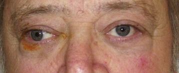
Cranial nerve III palsy is more common in patients older than 60 years and in those with diabetes or hypertension (Fig. 95.1).
![image](/wp-content/uploads/2016/06/B9781437735482000951_f095-001-9781437735482.jpg)
Fig. 95.1 This 60-year-old man had diabetes mellitus, hypertension, coronary artery disease, chronic renal failure, and multiple myeloma. He sought medical care because of double vision (he described the images as “a little side by side but mostly up and down”), diplopia, ptosis, and papillary sparing. Findings on laboratory tests, magnetic resonance imaging, and magnetic resonance angiography were negative. The patient was evaluated by a neurologist and an ophthalmologist, and diabetic cranial nerve palsy was ultimately diagnosed. He was given an eye patch and scheduled for ophthalmologic follow-up.
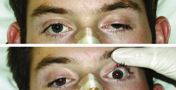
Patients with herniation syndromes will have a history of trauma (Fig. 95.2), tumor, or other neurologic findings.7
![image](/wp-content/uploads/2016/06/B9781437735482000951_f095-002-9781437735482.jpg)
Fig. 95.2 Ptosis and mydriasis suggest a cranial nerve III palsy. The appearance of these signs after a crush injury indicates that a skull fracture is impinging on the nerve canal.
(Reproduced with permission from Baker C, Cannon J. Images in clinical medicine. Traumatic cranial nerve palsy. N Engl J Med 2005;353:1955.)
Pain associated with unilateral mydriasis should alert the emergency physician (EP) to look for an aneurysm involving the terminal internal carotid artery. Computed tomographic angiography is more reliable than magnetic resonance angiography.8
Patients with an abscess or cavernous sinus thrombosis may have headaches, altered mental status, and seizures. This diagnosis should be considered in patients with signs and symptoms in the contralateral eye, previous sinus or midface infection, fever, chemosis, eyelid or periorbital edema, and exophthalmos. Extension of internal carotid artery dissection intracranially into the cavernous sinus can result in third, fourth, and sixth cranial nerve palsies.9
Cranial Nerve IV (Trochlear Nerve)
Anatomy
The trochlear nerve innervates the superior oblique muscle of the eye and causes inward rotation and downward and lateral movement of the globe. It is the smallest cranial nerve but has the longest intracranial course.
Presenting Signs and Symptoms
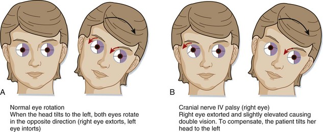
Patients with a fourth cranial nerve palsy have double vision exacerbated by looking downward. The classic complaint is difficulty going down stairs. Most commonly, a history of trauma is reported. On physical examination the patient may unconsciously tilt the head away from the affected side (Fig. 95.3). Etiologic mechanisms are similar to those for the third cranial nerve and include inflammatory processes, trauma, and vascular causes.10
Treatment
Treatment of isolated fourth nerve palsy is generally conservative, and the patient should be referred to neurology or neurosurgery as appropriate.10 CT, MRI, and neurology consultation are warranted if multiple cranial nerves are involved.
Cranial Nerve V (Trigeminal Nerve)
Anatomy
The trigeminal nerve is a mixed motor and sensory nerve. It provides motor innervation to the muscles of mastication, as well as sensation from the face, scalp, conjunctiva, globe, mucous membranes of the sinuses, tongue, teeth, and part of the external tympanic membrane.
The trigeminal sensory ganglion is located in the middle cranial fossa and branches into three divisions: the ophthalmic nerve (V1), the maxillary nerve (V2), and the mandibular nerve (V3).
Presenting Signs and Symptoms
Patients with trigeminal nerve dysfunction have either sensory or motor deficits. Sensory dysfunctions include paroxysmal pain, paresthesias (abnormal sensations such as burning, pricking, tickling, or tingling), dysesthesias (disagreeable, unpleasant, or painful sensations produced by ordinary stimuli), and anesthesia (loss of sensation). The motor dysfunction is usually described as difficulty chewing and difficulty swallowing.
Peripheral lesions cause loss of sensation or pain in only one division. Positive findings in two or more divisions (e.g., loss of light touch in one division and loss of sensitivity to pain, temperature, or pinprick in another division) should raise suspicion for a central cause.
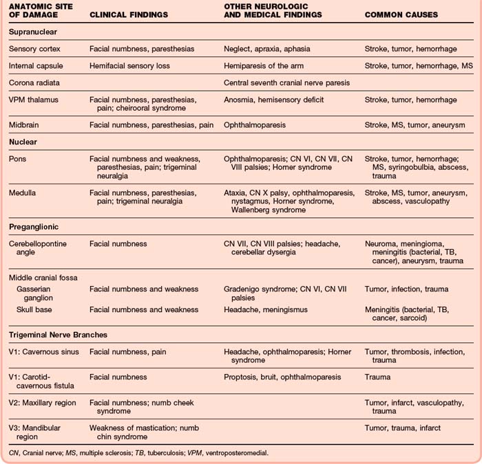
The presence of associated cranial nerve deficits (III, IV, IV, or any combination of these nerves) suggests cavernous sinus involvement. In the setting of trauma, if a bruit over the orbit can be detected, a carotid–cavernous sinus fistula may be present. Associated involvement of cranial nerve VII or VIII or gait ataxia should raise suspicion for a cerebellopontine angle or lateral pontine tumor (Table 95.1).
Associated Horner syndrome may indicate a cervical or lateral brainstem lesion.
The main categories of trigeminal nerve dysfunction are trigeminal neuralgia and trigeminal neuropathy. A sudden onset of symptoms should raise suspicion for a vascular, traumatic, or demyelinating cause, whereas a more indolent course suggests tumor or inflammation (Table 95.2).
Table 95.2 Selected Specific Causes Associated with Trigeminal Nerve Disorders
ETIOLOGIC CATEGORY | SELECTED SPECIFIC CAUSES |
---|---|
Structural Disorders | |
Developmental | Brainstem vascular loop, syringobulbia |
Degenerative and compressive | Paget disease |
Hereditary and Degenerative Disorders | |
Chromosomal abnormalities, neurocutaneous disorders | Hereditary sensorimotor neuropathy type I, neurofibromatosis (schwannoma) |
Degenerative motor, sensory, and autonomic disorders | Amyotrophic lateral sclerosis |
Acquired Metabolic and Nutritional Disorders | |
Endogenous metabolic disorders | Diabetes |
Exogenous disorders (toxins, illicit drugs) | Trichloroethylene, trichloroacetic acid |
Nutritional deficiencies, syndromes associated with alcoholism | Thiamine, folate, vitamin B12, pyridoxine, pantothenic acid, vitamin A deficiencies |
Infectious Disorders | |
Viral infections | Herpes zoster, unknown |
Nonviral infections | Bacteria, tuberculous meningitis, brain abscess, Gradenigo syndrome, leprosy, cavernous sinus thrombosis |
HIV infection, AIDS | Opportunistic infection; abscess, herpes zosterStroke, hemorrhage, aneurysm |
Neurovascular Disorders | |
Neoplastic Disorders | |
Primary neurologic tumors | Glial tumors, meningioma, schwannoma |
Metastatic neoplasms, paraneoplastic syndromes | Lung, breast; lymphoma, carcinomatous meningitis |
Demyelinating Disorders | |
Central nervous system disorders | Multiple sclerosis, acute demyelinating encephalomyelitis |
Peripheral nervous system disorders | Guillain-Barré syndrome, chronic inflammatory demyelinating polyneuropathyTolosa-Hunt syndrome, sarcoidosis, lupus, orbital pseudotumor |
Autoimmune and Inflammatory Disorders | |
Traumatic Disorders | Carotid-cavernous fistula, cavernous sinus thrombosis, maxillary/mandibular injury |
Epilepsy | Focal seizures |
Headache and Facial Pain | Raeder neuralgia, cluster headache |
Drug-Induced and Iatrogenic Neurologic Disorders | Orbital, facial, dental surgery |
AIDS, Acquired immunodeficiency syndrome; HIV, human immunodeficiency virus.
From Goetz CG, editor. Textbook of clinical neurology. 2nd ed. Philadelphia: Saunders; 2003.
Trigeminal Neuropathy
Causes include compression by an extrinsic mass, trauma, and vascular, inflammatory, or demyelinating disorders.
Symptoms include neuralgia or paresthesia (or both) involving half of the face. Unlike trigeminal neuralgia, the pain with trigeminal neuropathy is more constant. Loss of the corneal reflex is evident. The patient’s mouth may become more oval and oblique in appearance, and because of loss of masseter muscle strength, the chin may be deviated toward the affected side.
Until proved otherwise, neuropathies of cranial nerve V, the chin (numb chin; V3), and the suborbital region (numb cheek) should be presumed to be due to malignancies.11
![](https://clinicalpub.com/wp-content/uploads/2023/09/256.png)
Full access? Get Clinical Tree
![](https://videdental.com/wp-content/uploads/2023/09/appstore.png)
![](https://videdental.com/wp-content/uploads/2023/09/google-play.png)