Commonly Used Monitoring Techniques
The purpose of monitoring equipment is to augment the situational awareness of the anesthesiologist so that clinical problems can be recognized and addressed in a timely manner and to guide treatment. (Connor CW. Commonly used monitoring techniques. In: Barash PG, Cullen BF, Stoelting RK, Cahalan MK, Ortega R, Stock MC, eds. Clinical Anesthesia. Philadelphia: Lippincott Williams & Wilkins; 2013:699–722). Current American Society of Anesthesiologists (ASA) monitoring standards focuses attention on continually evaluating the patient’s oxygenation, ventilation, circulation, and temperature.
I. Monitoring Inspired Oxygen Concentratoin
Principles of Operation. Three main types of oxygen analyzer are seen in clinical practice: paramagnetic oxygen analyzers, galvanic cell analyzers, and polarographic oxygen analyzers (commonly used in anesthesia monitoring and are important components of gas machine oxygen analyzers, blood gas analyzers, and transcutaneous oxygen analyzers).
Proper Use and Interpretation.
The concentration of oxygen in the anesthetic circuit must be measured. (Anesthesia machine manufacturers place oxygen sensors on the inspired limb of the anesthesia circuit to detect and alarm in the event that hypoxic gas mixtures are delivered to the patient.)
As part of the preoperative checkout of the anesthesia machine, the clinician must confirm that the alarm limits of the inspired oxygen analyzer are set appropriately to alert to the presence of hypoxic mixtures.
Inspired oxygen alarms cannot be relied on to detect disconnection of the circuit.
Indications. The ASA Standards for Basic Anesthesia Monitoring states: “During every administration of general anesthesia using an anesthesia machine, the concentration of oxygen in the patient breathing system shall be measured by an oxygen analyzer with a low oxygen concentration limit alarm in use.” Careful monitoring of the inspired oxygen concentration is of particular significance during low-flow anesthesia in which the anesthesiologist attempts to minimize the fresh gas flow to the amount of oxygen necessary to replace the patient’s metabolic utilization.
Contraindications. The requirement to monitor inspired oxygen concentration may be waived by the responsible anesthesiologist under extenuating circumstances.
Common Problems and Limitations. Adequate inspiratory oxygen concentration does not guarantee adequate arterial oxygen concentration. (Additional monitoring for blood oxygenation includes adequate lighting and exposure to assess the patient’s color by direct observation.)
II. Monitoring of Arterial Oxygenation By Pulse Oximetry
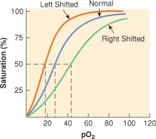
Principles of Operation. Pulse oximeters measure pulse rate and estimate the oxygen saturation of hemoglobin (SpO2) on a noninvasive, continuous basis. The oxygen saturation of hemoglobin (as a percentage) is related to the oxygen tension (as a partial pressure, mm Hg) by the oxyhemoglobin dissociation curve (Fig. 25-1).
Proper Use and Interpretation
At a constant light intensity and hemoglobin concentration, the intensity of light transmitted through a tissue is a logarithmic function of the oxygen saturation of hemoglobin.
The absence of a pulsatile waveform during extreme hypothermia or hypoperfusion can limit the ability of a pulse oximeter to calculate the Spo2.
The Spo2 measured by pulse oximetry is not the same as the arterial saturation (Sao2) measured by a laboratory co-oximeter.
In clinical circumstances when other hemoglobin moieties are present, the Spo2 measurement may not correlate with the actual Sao2 reported by the blood gas laboratory.
Increases in MetHb produce an underestimation when Spo2 is >70% and an overestimation when Spo2 is <70%.
COHb produces artificially high results.
Proper Use and Interpretation. Factors that may be present during pulse oximetry that affect its accuracy and reliability include dyshemoglobins, dyes (methylene blue, indocyanine green, and indigo carmine), nail polish, ambient light, light-emitting diode variability, motion artifact, and background noise. Electrocautery can interfere with pulse oximetry if the radiofrequency emissions are sensed by the photodetector.
Indications. Pulse oximetry has been used in all patient age groups to detect and prevent hypoxemia. Quantitative assessment of arterial oxygen saturation is mandated by the ASA monitoring standards.
Contraindications. There are no clinical contraindications to monitoring arterial oxygen saturation with pulse oximetry.
Common Problems and Limitations
Arterial oxygen monitors do not ensure adequacy of oxygen delivery to or utilization by peripheral tissues and should not be considered a replacement for arterial blood gas measurements or mixed central venous oxygen saturation.
Unless the patient in breathing room air, pulse oximetry is a poor indicator of adequate ventilation. (Patients who have been breathing 100% FiO2 may be apneic for several minutes before desaturation is detected by the pulse oximeter.) After the PAO2 has fallen sufficiently to cause a detectable decrease in SpO2, further desaturation may occur precipitously when the steep part of the oxyhemoglobin dissociation curve is reached.
III. Monitoring of Expired Gases
Principles of Operation. The patient’s expired gas is likely to be composed of a mixture of oxygen (O2), nitrogen (N2), carbon dioxide (CO2), and anesthetic gases (N2O and halogenated agents). Infrared absorption spectrophotometry (IRAS) gas analysis devices by transmitting light through a pure sample of a gas over the range of infrared frequencies creates a unique infra-red spectrum (like a fingerprint) for that gas.
Proper Use and Interpretation. Expired gas analysis allows detection of critical events (Table 25-1).
Interpretation of Inspired and Expired Carbon Dioxide Concentrations
Table 25-1 Detection of Critical Events by Implementing gas Analysis
Event
Gas Measured by Analyzer
Error in gas delivery
O2, N2, CO2, agent analysis
Anesthesia machine malfunction
O2, N2, CO2, agent
Disconnection
CO2, O2, agent analysis
Vaporizer malfunction or contamination
Agent analysis
Anesthesia circuit leaks
N2, CO2 analysis
Endotracheal cuff leaks
N2, CO2
Poor mask or LMA fit
N2, CO2
Hypoventilation
CO2 analysis
Malignant hyperthermia
CO2
Airway obstruction
CO2
Air embolism
CO2, N2
Circuit hypoxia
O2 analysis
Vaporizer overdose
Agent analysis
LMA = laryngeal mask airway.

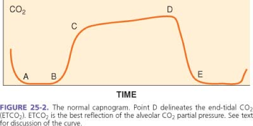
Figure 25-2. The normal capnogram. Point D delineates the end-tidal CO2 (ETCO2). ETCO2 is the best reflection of the alveolar CO2 partial pressure. See text for discussion of the curve.
Capnometry is the measurement and numeric representation of the CO2 concentration during inspiration and expiration.
A capnogram is a continuous concentration–time display of the CO2 concentration sampled at a patient’s airway during ventilation (divided into four distinct phases) (Fig. 25-2).
The first phase (A–B) represents the initial stage of expiration. Gas sampled during this phase occupies the anatomic dead space and is normally devoid of CO2.
At point B, CO2-containing gas presents itself at the sampling site, and a sharp upstroke (B–C) is seen in the capnogram.
Phase C to D represents the alveolar or expiratory plateau. At this phase of the capnogram, alveolar gas is being sampled. Normally, this part of the waveform is almost horizontal. However, when ventilation and perfusion are mismatched, Phases C to D may take an upward slope.
Point D is the highest CO2 value and is called the end-tidal CO2 (ETCO2). ETCO2 is the best reflection of the alveolar CO2 (PACO2).
Unless Rebreathing of CO2 occurs, the baseline approaches zero.
The ETCO2–PACO2 gradient typically is around 5 mm Hg during routine general anesthesia in otherwise healthy, supine patients.
Capnography is an essential element in determining the appropriate placement of endotracheal tubes.
The presence of a stable ETCO2 for three successive breaths indicates that the tube is not in the esophagus.
A continuous, stable CO2 waveform ensures the presence of alveolar ventilation but does not necessarily indicate that the endotracheal tube is properly positioned in the trachea. (Endobronchial intubation cannot be ruled out until breath sounds are auscultated bilaterally.)
A sudden drop in ETCO2 to near zero followed by the absence of a CO2 waveform heralds a potentially life-threatening problem that could indicate malposition of an endotracheal tube into the pharynx or esophagus, sudden severe hypotension, massive pulmonary embolism, a cardiac arrest, or a disconnection or disruption of sampling lines.
Gradual reductions in ETCO may reflect decreases in PaCO2 that occur when there exists an imbalance between minute ventilation and metabolic rate (CO2 production), as commonly occurs during anesthesia at a fixed minute ventilation.
Interpretation of Inspired and Expired Anesthetic Gas Concentrations. Monitoring the concentration of expired anesthetic gases (minimum alveolar concentration) assists in titrating those gases to the clinical circumstances of the patient.
Indications. Capnography is the standard of care for monitoring the adequacy of ventilation in patients receiving general anesthesia and during procedures performed while the patient is under moderate or deep sedation.
Contraindications. There are no contraindications to the use of capnography, provided that the data obtained are evaluated in the context of the patient’s clinical circumstances.
Common Problems and Limitations
Stay updated, free articles. Join our Telegram channel

Full access? Get Clinical Tree


