Common Emergency Procedures
Types of transfer/retrieval
Interhospital transfer (e.g. because the patient requires a specialist investigation or intervention; there is a lack of critical care beds in the referring hospital; to repatriate the patient).
2° retrieval (an interhospital transfer using a specialist team from another healthcare facility to retrieve the patient).
Decision to transfer
Is the transfer necessary?
Is further stabilization required; will further stabilization delay urgent treatment (e.g. surgical decompression of intracranial haematomas)?1
Is everything that might be needed to treat any reasonably predictable complication available to accompany the patient?
Are the correct staff, equipment, and transfer apparatus available?
Organization of transfer
Transfers should be organized using the ACCEPT format.
Assessment:
Review the history and assess the need for transfer
Clinically reassess the patient (following A, B, C, D, E, format)
Control:
Identify team members and allocate roles and tasks
Communication:
Ensure relatives and clinicians are aware of the transfer
For interhospital transfers, telephone the receiving unit before leaving
Evaluation:
Ensure transfer is appropriate, and decide the urgency of transfer
Decide what transfer vehicle and escort requirements are needed
Air transfer may be appropriate for long distances, or where speed is required, but may worsen hypoxia
Preparation:
Ensure the patient is appropriately ‘packaged’
Check equipment and reserves of O2, drugs, and battery life
Ensure all necessary notes and imaging are packaged
Transportation:
Confirm the route and the final destination
Continue monitoring during transit
Recheck the patient just before leaving, and just before arriving
Ensure handover of crucial information
Stabilization and preparation prior to transfer
Most procedures are difficult to undertake en route; if there is a probability of deterioration then anticipate and prepare for it.
Reassess the patient’s history, treatment, and examination findings.
Use a checklist wherever possible.
Airway
Consider endotracheal intubation if the airway may obstruct (especially in facial burns or trauma, or where GCS may drop to <8).
Confirm the ETT position and ensure it is secured firmly in place.
Decide whether C-spine precautions are required for transfer.
Some centres replace ETT cuff air with saline prior to air transport.
Breathing
If oxygenation/ventilation is poor consider elective intubation and mechanical ventilation prior to transfer:
NIV is rarely possible to deliver during transport
In ventilated patients with refractory hypoxia it may be possible to use portable ECMO for the transfer, this is provided by specialist centres.
Establish the patient on the transport ventilator prior to transfer and check ABGs:
Transport ventilators often only produce basic levels of respiratory support
Check for any chest problem, review CXR for evidence of pneumothoraces (difficult to treat in transit; may expand during air transfer)
Consider prophylactic chest drains in cases of chest trauma
Fluid-locked chest drains may be replaced with Heimlich valves
Circulation
Avoid transferring hypotensive patients where possible.
Ensure a minimum of 2 secure, working large-bore (16-G cannulae).
Control haemorrhage and fluid resuscitate.
If required, establish on inotropes/vasopressors prior to transfer:
Avoid using high concentration inotrope solutions where possible (>16 mg adrenaline/noradrenaline in 50 ml) as low volume siphoning from syringe drivers may occur
An invasive arterial line is preferable for BP monitoring:
NIBP monitoring is possible during road and air transport, but is less reliable and uses more battery power
If a PAFC is in situ and the waveform cannot be monitored in transit, consider withdrawing it to the RA to avoid inadvertent ‘wedging’.
Where arrhythmias are a problem place adhesive defibrillator pads on the patient prior to ‘wrapping’.
Stabilize long-bone/pelvic fractures.
Neurological
Other
Check available equipment for transfer:
Assemble and check critical pieces (e.g. self-inflating bags)
‘Mummy-wrap’ the patient with blankets (strap in all lines/tubes).
Ensure pressure point care and eye protection.
Remove/stop any unnecessary equipment or infusions.
If using air travel consider inserting an NGT and splitting plaster casts.
Consider the mode of transfer (ground versus air):
Air transfers may be high risk for patients with high O2 requirements, recent altitude sickness, recent diving injury, pneumothorax, bullae, pneumoperitoneum, or pneumocranium
Do they really need to fly; can the aircraft be pressurized to ground level equivalent?
Discuss morbidly obese patients with ambulance control as specialist bariatric equipment may be required.
Avoid using transfer oxygen/batteries until the last minute.
Communication and documentation
All notes, X-rays, CT scans, and blood results should be taken.
A patient summary sheet should be prepared where possible.
Ensure that all parties involved are aware of the transfer: the receiving hospital/unit, the patient/relatives, ambulance control:
Confirm the destination and obtain directions if appropriate
Consider a police escort for ‘life or death’ transfers
Transfer practices
‘Intensive care’ monitoring and management should be continued throughout the transfer process.
A minimum of 2 staff should accompany the patient, 1 of whom should be trained in advanced airway techniques for any patient who is, or may require to be, intubated; both staff should be familiar with transfer equipment.
Phone the receiving unit immediately prior to transfer.
Before leaving review the patient’s condition for a final time and check all the drugs and equipment needed are going with the patient.
Take money and an emergency phone with you (and the telephone numbers of the transferring and receiving units).
Management
Allocate any task-specific roles early (e.g. who is responsible for monitoring specialist equipment such as ICP monitoring).
Airway
Breathing
Mechanically ventilated patients often need to be sedated and given neuromuscular blocking drugs to facilitate safe transfer.
Transport ventilators are often less efficient, necessitating ↑ventilation pressures or higher FiO2, especially during air transport.
Special attention needs to be paid to chest injuries:
Do not clamp chest drains
Where pneumothoraces occur in mechanically ventilated patients during air/road transfers consider doing a mid-axillary line blunt-dissection thoracocentesis without inserting a chest drain (air should be released, the lung should be palpable confirming re-expansion, a sterile chest drain may be inserted upon arrival)
Calculate the volume of O2 required for the transfer:
Switch to ambulance/aircraft O2 supply as soon as appropriate
Circulation and neurological
Where possible keep infuser pumps at the same level as the patient (some pumps siphon).
Vibration in aircraft (particularly rotary wing) may interfere with gravity IV infusions, pressure infusers should be available.
Invasive BP monitoring is often more reliable than non-invasive techniques during transfer.
Infusion pumps, monitors and any associated transfer equipment must be attached to either the transfer trolley or the vehicle during transfers (to avoid them becoming projectiles in crash situations).
Maintain access to pupils (for assessment).
Environment
Keep the ambulance/aircraft warm.
Keep all monitoring within view.
Ensure IV access is always within reach.
Movement
Perform a visual sweep prior to the patient moving from any location to ensure lines, NGTs, urinary catheters are not going to catch.
Always ensure that the team are prepared and know exactly how (i.e. log roll, slowly) and which way the patient is going to be moved:
A recognized ‘count’ is: ‘ready, steady, slide/roll’
Secure the patient to any transfer trolley (cot-sides up, safety belts attached) prior to moving trolley; ensure trolley is secured to ambulance/aircraft prior movement.
Transport vehicles
Once inside a vehicle secure all equipment.
Staff should be securely seated during travel.
If possible the head and one side of the patient should be accessible.
Consider providing antiemetics to the patient (and staff).
Special considerations for aircraft:
Never approach/enter an aircraft without permission from the pilot or load-master; follow their instructions at all times
Never approach the rear of a rotary wing aircraft with a tail rotor
Consider providing ear-defenders, or other protection, to the patient during air transfers
‘Hot’ unloads are rarely necessary, full engine shutdown should normally be allowed to happen before departure from rotary wing aircraft; if a ‘hot’ unload is required make sure everyone knows their roles and responsibilities prior to landing (and that ground crew are aware)
Defibrillation in aircraft: most modern aircraft are defibrillationsafe, check first
Take-off, landing, and banking will increase physiological stresses (hypotension or raised ICP may be temporarily worsen); infusion pumps above or below the patient may have slightly altered rates
Air expansion at altitude:
Air-filled splints should be opened, plaster casts may need splitting, NG decompression may be required
Some guidelines recommend inflating endotracheal and catheter cuffs with saline rather than air (alternatively monitor pressures)
Air bubbles in invasive monitoring sets will expand and dampen pressure monitoring traces
Documentation
Take all relevant patient documents with you.
Document the patient’s condition during the transfer.
Perform a detailed handover of the patient to the receiving team.
Transfer equipment
Ensure you are familiar with available equipment
When retrieving patients assume that no equipment will be available: take your own
Use dedicated trolleys for interhospital transfers where available
Where transfer may be weight-limited (e.g. in rotary wing aircraft)—discuss any requirements with the crew.
Airway and ventilation
Guedel and nasopharyngeal airways (assorted sizes).
ETTs (assorted sizes); laryngoscopes (spare bulbs and battery); intubating stylet; lubricating gel; Magill’s forceps; tape for securing tracheal tube; sterile scissors; stethoscope; laryngeal mask airways (assorted sizes).
Portable suction; Yankauer sucker; suction catheters (assorted sizes); NG tubes (assorted sizes) and drainage bag.
Self-inflating bag and mask with O2 reservoir; high-flow breathing circuit; chest drains (assorted sizes); Heimlich flutter valves.
Ventilators: check prior to use; spare valves for portable ventilator.
Oxygen
Calculate O2 requirements for trip, ensure that 2-3 hours of ‘back-up’ O2 is available (discuss with ambulance crew the O2 they have available) (Table 17.1).
Many ventilators display their O2 consumption.
Circulation and drugs
Syringes (assorted sizes); needles (assorted sizes); alcohol wipes; IV cannulae (assorted sizes); arterial cannulae (assorted sizes); central venous cannulae; IV fluids; infusion sets/extensions; 3-way taps; dressings/tape; pressure infusers.
Emergency drugs for cardiac arrest and intubation/reintubation should be available; spare infusions should be prepared for any inotropes, sedatives, or muscle relaxants; predictable emergency drugs should also be available (e.g. anti-seizure medication); spare IV fluid should be available.
Infusion pumps should be checked (and spares taken where transfer will be long, or an infusion is critical); a defibrillator may be required (ambulance crews may be able to provide this).
Other equipment
Consider the need for: blood, minor instrument/cut-down set, tracheostomy set.
Specialist protective clothing should be available for interhospital transfers or retrievals; individuals roles should be clearly identifiable.
Pitfalls/difficult situations
Most problems during transfer result from inadequate stabilization prior to departure, or failure to continue optimal treatment and monitoring during transfer.
1With the exception of 1° retrievals, and transfers for urgently required treatments, there will be sufficient time to fully stabilize the patient. A ‘scoop and run’ policy should only be adopted where there are insufficient resources available to resuscitate A, B, and C.
Further reading
Ahmed I, et al. Risk management during inter-hospital transfer of critically ill patients: making the journey safe. Emerg Med J 2008; 25: 502-5.
Association of Anaesthetists of Great Britain and Ireland. Interhospital transfer, AAGBI safety guideline. London: AAGBI, 2009.
Intensive Care Society. Guidelines for the transport of the critically ill adult. London: Intensive Care Society, 2011.
Macartney I, et al. Transfer of the critically ill adult patient. Br J Anaesth CEPD Rev 2001; 1(1): 12-15.
Rapid sequence endotracheal intubation (RSI) is a complex skill, which should not be attempted without training. Even if you are not competent to perform the procedure you may be called upon to assist or prepare the equipment.
Indications for endotracheal intubation
To protect the airway
From risk of aspiration (e.g. blood/vomit).
From risk of airway obstruction.
Where sedation/anaesthesia is required to allow assessment or treatment (particularly in agitated or combative patients).
To permit mechanical ventilation
Apnoea or bradypnoea.
Hypoxia or inadequate respiratory effort.
Hypercapnia or requirement for hyperventilation.
Cardiovascular instability, to optimize oxygen supply/demand.
Indications for rapid sequence intubation
RSI is the preferred technique for endotracheal intubation where there is a risk of aspiration:
Aspiration of gastric contents is strongly associated with unstarved patients, pregnancy, oesophageal disease, obesity, ileus/bowel obstruction.
Contraindications
When attempted as an absolute emergency (i.e. impending or actual cardiac arrest) there are no contraindications to attempting endotracheal intubation.
In all other situations (i.e. urgent or semi-elective endotracheal intubation) the operator must be experienced in the technique and the likelihood of the patient being difficult to intubate must be assessed ( p.24) and appropriate preparations made.
Suxamethonium should not be used in patients with a known allergy to suxamethonium, a history of MH, hyperkalaemia, recent burns (>2 days, <18 months), or paralysis.
Equipment
One assistant (preferably more) is required to monitor the patient, administer drugs, pass equipment to the operator, and apply cricoid pressure (allocate roles beforehand, in case of difficult intubation).
A range of facemasks and a means of ventilating (i.e. self-inflating bag).
Cuffed ETT; as a rough guide size 8 for adult females, size 9 for males:
Range of cuffed ETTs (sizes 6.5-10)
Spare ETTs should be prepared in the sizes above and below the chosen size
ETTs may be pre-cut to a length of 26-28 cm; do not cut ETTs if oro-facial swelling is likely (e.g. burns)
Lubricant gel (to facilitate passage of ETT through vocal cords).
10-ml syringe (for inflating ETT cuff).
Laryngoscope (plus spare) with Mackintosh blades size 3 and 4.
Gum elastic bougie, stylet.
Tape or tie (to secure ETT post-intubation).
Stethoscope.
Yankauer sucker, suction catheters and suction apparatus, and a bed/trolley that can tilt head down if possible.
Magill forceps.
ETCO2 monitoring.
Intubation drugs (e.g. ketamine, thiopental, or propofol; suxamethonium or rocuronium).
Emergency drugs, immediately available:
Resuscitation drugs (e.g. metaraminol, atropine, adrenaline)
Failed intubation equipment, readily accessible:
Microlaryngel tubes (size 5-6)
Special laryngoscopes (e.g. McCoy, short-handled, polio blade)
LMAs/supraglottic airway devices (sizes 3, 4, 5) (intubating LMA if available)
Airway exchange catheters
Intubating fibreoptic scope
Berman airways
Emergency cricothyroidotomy kit
Jet ventilator
Consider having an NGT available.
Anatomical landmarks
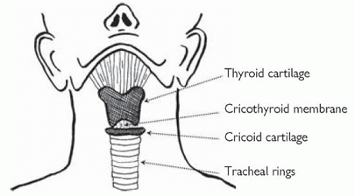
The cricoid cartilage may be identified immediately inferior to the thyroid cartilage; it feels like a wedding ring below the skin (Fig. 17.1).
Technique
Briefly assess the patient’s airway (see p.24); and check any pertinent history (e.g. allergies, previous anaesthetic reactions).
Check equipment (suction, laryngoscope bulb, ETT cuff).
Mentally prepare a plan A, B, and C in case of failed intubation (see p.25), ensure staff are informed of key aspects.
Establish IV access.
Aspirate NGT if one is in place.
Ensure the patient is positioned with their head on a pillow (often removed during CPR) so that the head is raised just above the shoulders.
Attach all monitoring; check that the bed/trolley head tilt mechanism is working.
Head positioning is not possible where trauma is suspected; in such cases an assistant is required to apply manual in-line stabilization to the head. The anterior aspect of the hard collar can then be undone or removed prior to attempting tracheal intubation. Where possible a bougie should be used for trauma intubations in order to minimize the degree of neck movement required.
Pre-oxygenate with 100% O2 for 3-5 minutes if possible, using a facemask and ventilating circuit (gently assisting ventilation is best avoided if possible, but may be necessary in critically ill patients).
Remove false teeth.
Have suction turned on and within reach.
Administer induction agents over 20-30 seconds, any of:
Thiopental IV 2-5 mg/kg
Propofol IV 1-5 mg/kg
Ketamine IV 1-3 mg/kg
Thiopental and propofol commonly cause hypotension requiring fluid and/or inotropic support; in cardiovascularly unstable patients consider using ketamine (may cause hypertension, tachycardia or raised ICP)
Apply cricoid pressure (do not release until ETT is in position, ETCO2 is present and the cuff is inflated1).
Immediately administer suxamethonium IV 1-2 mg/kg (where suxamethonium is contraindicated use rocuronium IV 0.6-1 mg/kg):
Suxamethonium should cause fasciculation within 15-30 seconds (not always observed).
After 30 seconds (or when fasciculations cease, or 60 seconds if using rocuronium), holding the laryngoscope in your left hand (close to the angle between the handle and blade), insert the blade into the right-hand side of the patients’ mouth:
Bring the laryngoscope back to the midline, pushing the tongue aside beneath it; advance forwards until the tip of the laryngoscope is in the vallecula space, then lift vertically until the vocal cords are seen (do not lever scope backwards onto teeth)
Pass a size 8-9 ETT through the cords.
Inflate the ETT cuff and attempt ventilation.
Confirm correct position of the ETT within the trachea by:
Obvious chest movement
Presence of misting/clearing of the ETT/catheter mount
Bilateral chest sounds on auscultation (listen to the chest in the mid-axillary line on both sides)
Absence of ‘bubbling’ sounds over the stomach on auscultation
If intubation is successful and the patient stable consider inserting an NGT or OGT.
Commence a sedative infusion.
Consider a bolus dose of non-depolarizing muscle relaxant (unless rocuronium already used).
Stay updated, free articles. Join our Telegram channel

Full access? Get Clinical Tree


