Category
Causes
Hernia
Indirect inguinal hernia
Direct inguinal hernia
Femoral hernia
Hip joint injury
Avulsion fracture
Stress fracture
Labral tear
Loose bodies
Degenerative joint disease
Femoroacetabular impingement
Legg–Calvé–Perthes disease
Slipped capital femoral epiphysis
Osteonecrosis
Athletic injuries
Sportsmans hernia (inguinal disruption)
Osteitis pubis
Adductor muscle strain
Genitourinary
Ectopic pregnancy
Round ligament pain
Endometriosis
Ovarian cyst
Ovarian torsion
Varicoceles
Prostatitis
Orchialgia
Urinary tract infection
Gastrointestinal
Appendicitis
Diverticulitis
Inflammatory bowel disease
Irritable bowel syndrome
Adhesions
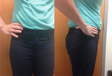
Particular attention should be paid to the acuity of the injury. In athletes, many acute groin injuries arise from strains of the adductor muscles or hip flexors and will resolve with conservative measures [5]. Chronic causes of groin pain are less likely to resolve and generally require more definitive treatments [5–8]. Schilders and colleagues classified chronic sources of groin pain in athletes into four separate categories: hip joint injury, osteitis pubis, adductor dysfunction, and sports hernias [9]. Patients with hip joint injuries will commonly indicate their pain with the C sign (Fig. 4.1), where the hand is cupped over the hip in the shape of the letter C with the ipsilateral index finger positioned over the groin and the thumb located proximal to the greater trochanter [10]. Patients with adductor injuries often complain of a pulling or tearing sensation in the groin with activity, while those with osteitis pubis note tenderness over the pubic symphysis. Patients with sports hernias typically complain of pain that is unilateral and burning or sharp in nature. The pain may radiate to a variety of locations, including the proximal thigh, lower back, lower abdomen, and downward to the scrotum as well [11]. Patients are generally able to sleep comfortably through the night, but upon awakening may experience extreme pain while attempting to get out of bed. Sudden movements, especially rotational or forceful activities such as sit-ups, cutting, and rapid acceleration or deceleration, will exacerbate these symptoms [5], while periods of rest will often relieve them, only to have them return upon resuming athletic activities [12].


Fig. 4.1.
In The “C Sign,” the hand is cupped over the hip in the shape of the letter C with the ipsilateral index finger positioned over the groin and the thumb located proximal to the greater trochanter.
Patients with inguinal hernias as their source of groin pain may experience a different set of symptomatology from those with sports hernias. The pain or discomfort associated with the inguinal hernia tends to be progressive over the course of the day and will be worse even in the evenings. Certain positions that increase intra-abdominal pressure, such as sitting, may exacerbate these symptoms, while lying supine may relieve them and return the hernia contents to their intra-abdominal location. Many patients will note increases in pain with forceful activities such as sneezing, coughing, and bowel movements, and some will reflexively hold their groin during these activities in order to mitigate these symptoms. Unlike patients with sports hernias, those with inguinal or femoral hernias will often notice a bulge and may associate their symptoms with a change in its size. All patients should be asked about any change in bowel or bladder habits to assess for any obstructive type symptoms and should also be asked about any history of incarceration of their hernias.
While traditional hernias, sports hernias , and other sport-related musculoskeletal injuries comprise the majority of causes for groin pain, there are many other congenital and acquired causes of groin pain that should be factored into one’s differential diagnosis. Congenital disorders associated with osteonecrosis of the hip, including slipped capital femoral epiphysis, congenital hip dysplasia, and Legg–Calvé–Perthes disease , may all be a source of groin or hip pain in adulthood [13]. Other causes of osteonecrosis such as chronic steroid use or alcohol abuse should also be assessed. Many genitourinary conditions may present with groin pain as well, and these should be discussed in detail, especially with sexually active women and women of childbearing age where the differential diagnosis should include conditions such as pelvic inflammatory disease, ectopic pregnancy, ovarian cysts, endometriosis, ovarian torsion, and round ligament pain.
An accurate and thorough history is the key initial step in the workup of all patients with groin pain. While the history alone may not confirm the diagnosis, it will help determine which physical exam maneuvers and ancillary studies will be most appropriate and helpful in determining the cause of the patients’ complaints.
Physical Exam
The physical examination should begin with vital signs, including accurate height and weight . Overweight and obese patients have a lower incidence of inguinal hernia formation compared to normal weight individuals [14, 15]. Routine physical examination of the thorax and abdomen should be performed, but the majority of the physical exam should be focused on the groin and the hip. The back, pelvis, groin, and upper thigh should be completely exposed in order to facilitate a thorough examination.
Examination should begin in the upright position with inspection. Palpation of the spine and paraspinal muscles should be done. Unilateral inguinal hernias will often be apparent as an asymmetric bulge in the inguinal area that may or may not extend into the scrotum in males. Most other pathologies will have no obvious findings on inspection alone. Palpation of the groin should also begin in the upright or standing position. Many inguinal hernias can be palpated simply by placing the hand over the inguinal canal and reducing any hernia contents into their intra-abdominal position. The patient is then asked to cough or to perform the Valsalva maneuver, and the hernia contents should slide past one’s fingers. If a hernia cannot be appreciated, the index finger can be placed into the inguinal canal by invaginating the scrotum in male patients. With a finger placed deep in the canal, hernias can again be appreciated with Valsalva or cough . Additionally, the inguinal occlusion test can be performed to determine if the hernia is direct or indirect [16]. With this maneuver, the hernia contents are reduced and manual pressure is applied over the presumed site of the deep inguinal ring. The patient then performs a Valsalva maneuver, and one can observe if the hernia appears with continued compression (direct) or only after release of the internal ring (indirect). While this maneuver may help to differentiate the types of inguinal hernia, its accuracy is relatively low and is not likely to alter the surgical intervention [17, 18]. If no hernia is appreciated, then the groin is similarly examined with the patient lying supine. If hernias still cannot be recognized, then ancillary imaging may be necessary or an alternative diagnosis should be entertained.
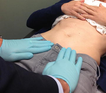
Patients with sports injuries, often suspected by the patient’s history, should undergo a sequential exam of the back, hip, and groin [19, 20]. For a general surgeon, the exams will in most cases be basic, but even a basic exam will help direct referral or image ordering. The spine and back should be palpated along the thoracic, lumbar, and sacral vertebrae. The paraspinal muscles should also be palpated, and the rare entity of thoracolumbar syndrome should be ruled out when this entity is suspected. The hip should then be examined with some simple maneuvers that examine hip rotation, extension, and flexion. The rest of the exam should focus on the groin, where a firm understanding of the musculoskeletal anatomy will help greatly in figuring out the precise cause of the athletic pubalgia. The rectus muscle insertion on the pubis should be examined with palpation and a sit-up or crunch maneuver while palpating the conjoint tendon Fig. 4.2 [11]. Reproducible pain in this area suggests rectus sheath or conjoined tendon pathology. The pubic tubercle should be palpated and pain with direct pressure can suggest osteitis pubis [21]. Finally, the leg muscles , specifically the adductors and abductors, can be examined by asking the patient to adduct and abduct against resistance and noting any reproducible symptoms [20]. The hip flexors (iliopsoas and rectus femoris) can also be tested at this point with leg flexion against resistance. If all is normal, yet the patient only has tenderness over the internal ring region of the canal without a palpable hernia, a “sports hernia” can then be suspected.


Fig. 4.2.
The examiner places pressure on both groins, while the patient actively sits up. Pain indicates a possible inguinal disruption injury.
There are a wide variety of physical exam maneuvers that can be employed to assess for hip joint injury as the cause of groin pain (Table 4.2) [1, 10, 22–25]. Range of motion, strength, and provocative maneuvers may all be necessary; however, the majority of these clinical tests have not been found to be of substantial quality to dictate clinical decision making [26]. The majority of these maneuvers are outside the scope of practice for most general surgeons, and if hip pathology is suspected, then early referral to Sports Medicine or Orthopedic Surgery is warranted, as radiographic studies will likely be necessary to make a firm diagnosis.
Maneuver | Examination procedure | Results | Possible diagnoses |
|---|---|---|---|
Dynamic internal rotary impingement | Patient is laid supine with the contralateral leg flexed beyond 90° at the hip. The ipsilateral hip is flexed to 90° and passively taken through a wide arc of adduction and internal rotation | Pain | Femoroacetabular impingement Labral tears |
Dynamic external rotary impingement | Patient is laid supine with the contralateral leg flexed beyond 90° at the hip. The ipsilateral hip is flexed to 90° and passively taken through a wide arc of abduction and external rotation
Stay updated, free articles. Join our Telegram channel
Full access? Get Clinical Tree
 Get Clinical Tree app for offline access

|


