Care of the Mechanically Ventilated Patient with a Tracheotomy: Introduction
Although Egyptian tablets depict use of tracheotomy for medical applications nearly 5600 years ago,1 initial descriptions of the procedure in Western literature did not appear until the middle of the sixteenth century.2 By 1718, “tracheotomy” became accepted terminology for the surgical technique that was then primarily used for relief of airway obstruction and removal of aspirated foreign bodies. Typically gruesome clinical results relegated tracheotomy to a reviled role in airway management and gained it a designation as the “scandal of surgery.”3 The diphtheria epidemics of the nineteenth century popularized tracheotomy.4 Tracheotomy did not become widely accepted, however, until 1909, when Chevalier Jackson standardized surgical techniques and decreased the operative mortality from 25% to less than 1%.5
Advances in tube design during the 1960s and 1970s and improved management techniques further promoted acceptance of tracheotomy for long-term airway access for critically ill, ventilator-dependent patients. The advent of percutaneous dilational tracheotomy (PDT) further widened the application of tracheotomy by allowing the procedure to be performed in the intensive care unit (ICU) by nonsurgeons.6 Up to 24% of patients undergoing mechanical ventilation and 6% of critically ill patients in general have a tracheotomy performed.6–12 In North Carolina, nearly a threefold increase in the application of tracheotomy for prolonged mechanical ventilation was observed from 1993 to 2002.9 Although only 7% of ventilated patients underwent tracheotomy in that study, they accounted for 22% of all mechanical ventilation patient charges.
Four indications exist for a tracheotomy in critically ill patients: (a) maintenance of airway patency for patients with functional or mechanical upper airway obstruction, (b) provision of airway access for suctioning retained airway secretions, (c) prevention or limitation of aspiration in patients with glottic dysfunction, and (d) management of patients who require long-term airway access for ventilator support.13 Acceptable outcomes from tracheotomy remain dependent on the skill of the operator who performs the procedure and expertise of interdisciplinary teams charged with managing patients from the critical care phase of their illnesses through transitions to hospital wards and long-term care facilities until successful decannulation occurs.14,15
Techniques of Surgical Airway Access
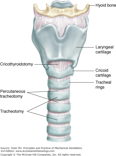
A standard surgical tracheotomy provides tracheal access through a temporary incisional tracheostoma between cartilaginous rings (Fig. 40-1). The stoma spontaneously closes after removal of the tracheotomy tube.
Standard surgical tracheotomy is a well-tolerated procedure with an operative mortality of less than 1% when performed as an elective procedure in stable, ventilator-dependent patients. In contrast, most,16 but not all,17 centers report a higher complication rate when surgical tracheotomy is performed as an emergency procedure. Some groups report successful emergency tracheotomy when performed on awake patients.18 Most centers, however, have replaced emergency surgical tracheotomy with specialized endotracheal intubation techniques to secure a difficult airway,19 emergency cricothyroidotomy,20 and emergency percutaneous techniques.21–27
Percutaneous tracheotomy refers to several techniques that insert a standard or modified tracheal airway with a Seldinger technique below the first or second tracheal rings using a device to cut and spread the trachea28 or a forceps or dilator technique to cannulate and dilate tracheal tissue between cartilaginous rings (see Fig. 40-1).29–31
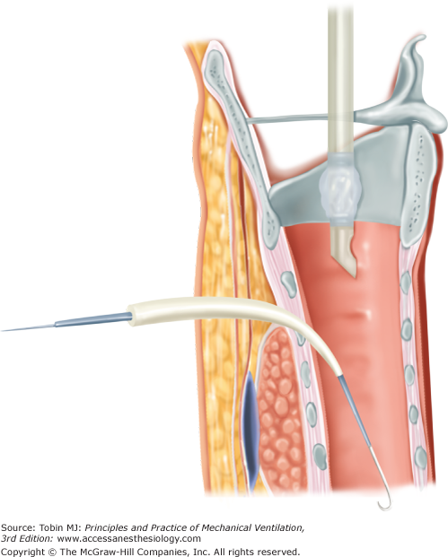
Ciaglia first described percutaneous dilatational tracheotomy (PDT) wherein a Seldinger technique allowed the insertion of sequential dilators to place a tracheotomy tube.30 The procedure has evolved from the use of sequential dilators to insertion of a single dilator that increases in caliber from tip to base where it matches the diameter of a tracheotomy tube (Ciaglia Blue Rhino, Cook Critical Care Inc., Bloomington, IN) (Fig. 40-2).32 The single-dilator Blue Rhino technique results in faster insertion times as compared with the sequential dilator technique,27,33,34 and a lower risk of posterior tracheal injury.27,35,36
Recently, the Ciaglia PDT has been further modified with use of a balloon dilator (Ciaglia Blue Dolphin, Cook Critical Care Inc., Bloomington, IN), which is placed across the anterior tracheal wall and inflated to dilate the stoma by radial force, thereby avoiding anterior–posterior compression of tracheal structures that can fracture cartilaginous rings. The procedure has not yet been established as being superior to the Blue Rhino technique.37,38
The Ciaglia PDT technique is the most commonly performed percutaneous tracheotomy procedure in the United States and is frequently used in Europe.39–41 Nonsurgeons can perform the procedure with low complication rates, with few patients requiring conversion to a surgical tracheotomy.42 Controversy remains, however, among otolaryngologists, with only 29% of otolaryngology residency programs teaching PDT; however, programs that do include training in PDT use the Ciaglia Blue Rhino technique and endorse its safety.43,44
Other techniques for percutaneous tracheotomy include a specialized guidewire dilating forceps with a groove that allows loading of a guidewire onto forceps, which dilate the trachea, thread the wire, and insert a tracheotomy tube by Seldinger technique (Portex guidewire dilating forceps kit, Sims, Inc, Philadelphia, PA).45 The guidewire dilating forceps technique provides quick, safe, and effective placement of a tracheotomy for critically ill patients.46–51 Guidewire dilating forceps require the same time to completion as the Ciaglia single-dilator method.52,53 Comparative studies have shown both higher,54 similar,46,52,53 and lower50 complication rates with the guidewire dilating forceps as compared with the Ciaglia Blue Rhino technique, with each having unique complications.55 Long-term follow-up studies demonstrate acceptable rates of tracheal stenosis and other chronic airway complications with guidewire dilating forceps,56–59 although one study of 208 patients observed a 38% incidence of a changed voice and a 12% incidence of persistent, severe cough. Patients with short, fat necks, conditions that prevent neck extension, an enlarged thyroid isthmus, a previous tracheotomy, coagulopathy, or anticoagulation therapy have successfully undergone the guidewire dilating forceps technique.60
Frova et al described a single-step technique (PercuTwist, Rusch, Kernen, Germany) that threads a screw-type dilator between tracheal rings to allow insertion of a specially designed tube.61 This technique causes less compression of the anterior tracheal wall than does the Ciaglia technique. Early experience with PercuTwist reported variable success with difficulties in learning the procedure and problems with damage to tracheal structures.34,62–65 More recent reports using bronchoscopic guidance compared PercuTwist with other percutaneous techniques and reported similar complication rates and speed of completion of the procedures.66–68
Fantoni proposed translaryngeal tracheotomy for placing a tracheotomy in a reverse direction from within the airway through the tracheotomy tract.69 The operator places a needle into the trachea between the second and third tracheal rings. With bronchoscope guidance, a guidewire is passed retrograde through the needle into a cuffed, rigid bronchoscope, which is then removed from the airway. A tracheotomy tube with a tapered proximal end is loaded onto the cephalad end of the guidewire and pulled through the airway and the needle tract. Comparative studies suggest that translaryngeal tracheotomy is equally effective and less traumatic than the Ciaglia PDT technique.70–73
All percutaneous techniques require a learning curve to gain skills for completing the procedures quickly and safely.74,75 Centers that emphasize close supervision of trainees note no differences in complication rates by seniority of operator.76 Because PDT is the primary technique used in most ICUs, it is the percutaneous technique discussed in the remainder of this chapter.
Contraindications to PDT have decreased with increasing experience with the procedure. Conservative relative contraindications have included age younger than 16 years, bleeding diatheses, severely calcified tracheal rings, and anatomic abnormalities, such as obesity and thyromegaly, that obscure landmarks.35,77–79 Greater experience with PDT, however, has allowed its application by experienced practitioners in patients with body mass indices equal to or greater than 27 kg/m280 and equal to or greater than 35 kg/m2.81 Preoperative ultrasonography allows selection of patients with anatomic abnormalities of the neck for successful PDT.82,83 Trauma and burn patients with known or suspected cervical injury, patients 5 to 6 days after anterior spinal fusion, and patients with previous tracheostomies have also undergone successful PDT.83–89 Studies report successful PDT for patients with thrombocytopenia,90,91 coagulopathies secondary to liver disease,92 and systemic heparinization.91,93 PDT has been performed successfully for patients with acute respiratory failure and dependence on high levels of positive end-expiratory pressure (mean: 16.6 cm H2O; range: 12 to 20 cm H2O positive end-expiratory pressure).94
Purported advantages of PDT as compared with standard surgical tracheotomy include speed of the procedure (less than 10 minutes), ICU placement without operating room transport, avoidance of general anesthesia, decreased cost, reduced operating room use, and limited personnel requirements that include a single operator, a bronchoscopist, a nurse, and a respiratory therapist.95,96 Such simplifications decrease costs,97 avoid complications from intrahospital transport, and eliminate dependency on the operating room schedule, thereby decreasing the time from the decision to place a tracheotomy to when it is actually performed.98 Some centers, however, obviate some of these benefits by performing standard surgical tracheotomy in the ICU.99–109 Standard surgical tracheotomy in the ICU has similar costs and complication rates comparable to PDT if performed under controlled circumstances with all of the resources that would be available in the operating room.108,109
Percutaneous tracheotomy has a potential advantage over standard surgical tracheotomy by dilating rather than incising cervical tissues thereby producing a tracheostoma that fits snugly around the tracheotomy tube.110 This tight fit has potential for improving anchoring of the tracheotomy tube, decreasing the incidence of inadvertent extubation (1.4% incidence),43 compressing blood vessels in the stoma tract thus decreasing postoperative bleeding (2% to 4% incidence),35,43,111 and diminishing the incidence of wound infections observed with conventionally placed surgical stomas.111 The smaller skin incision also may result in a better cosmetic appearance after extubation.111,112
Many observational studies and a smaller number of randomized, controlled trials73,86,95,96,98,104,111–119 have addressed the potential benefits of PDT by comparing the outcomes of the procedure with those of standard tracheotomy. These trials, however, are relatively small, often employ varying tracheotomy techniques, have considerable spectrum bias, usually exclude patients with relative complications to PDT, and measure heterogeneous outcomes with differing definitions between studies. Five meta-analyses have critically analyzed these studies and come to varying conclusions.109,120–123 The meta-analysis by Dulguerov et al concluded that PDT had a higher complication rate than standard tracheotomy, but the analysis suffered from methodological flaws (double counting) and included retrospective studies.109 The three most recent meta-analyses109,122,123 have the usual limitations from heterogeneity of primary studies, but conclude that PDT and surgical tracheotomy have comparable major complication rates. The analyses differ regarding relative rates of early postoperative complications, but Delaney et al performed a subgroup analysis by type of PDT and found a similar rate of postoperative bleeding for PDT as compared with surgical tracheotomy performed in the operating room.123 General conclusions from these analyses indicate that the choice of technique should be individualized by an interdisciplinary approach and PDT can be performed more quickly with similar outcomes as compared with surgical tracheotomy performed in the operating room or at the bedside in appropriately selected patients.109 A shorter time from the decision to perform tracheotomy to its actual completion may result from PDT.98,111 Prospective comparative data of surgical versus PDT for patients with underlying bleeding diatheses, neck anatomic abnormalities, and other risk factors for complications do not exist.124 Also, up to 7% of PDT procedures require conversion to surgical tracheotomy because of technical difficulties.122
Cricothyroidotomy, or more specifically surgical cricothyroidotomy, is a surgical technique for placing an airway through the cricothyroid space (see Fig. 40-1). The term has also been applied to percutaneous placement of a cannula by Seldinger technique (cannula or percutaneous cricothyroidotomy) or a needle (needle cricothyroidotomy) through the cricothyroid space to establish an emergency airway. Because of its simplicity and the superficial location of the cricothyroid space, cricothyroidotomy has become the preferred procedure for emergency airway placement for patients who cannot undergo translaryngeal intubation.125–128 After training on simulators or cadavers, operators can complete surgical or cannula cricothyroidotomy in less than 60 seconds.129–131 Some centers, however, prefer emergency PDT after failed intubation attempts.21–27 Up to 62% of patients have vertically oriented arteries and veins overlying the cricothyroid membrane, which can complicate cricothyroidotomy.132
Most intensivists do not employ cricothyroidotomy for elective, long-term airway access in critically ill patients because of concern for delayed airway damage, which has been reported to occur more commonly in patients with 6 to 7 days of prior translaryngeal intubation.133–137 More recent studies propose that cricothyroidotomy has a low complication rate and represents a reasonable option for critically ill patients with challenging neck anatomy.106,138,139 These studies were retrospective, however, and did not provide systematic patient follow-up to detect long-term airway complications.
Cricothyroidotomy presents potential long-term risks to vocalization after decannulation because the tube inserts through the cricothyroid membrane, which lies within 1 cm of the true vocal cords, and can thereby cause glottic and direct vocal cord scarring and also prevent anterior pivoting of the laryngeal cartilage, which is required to stretch the vocal cords.140,141 Up to 78% of patients treated with a cricothyroidotomy experience hoarseness.140–144 No studies, however, have compared the relative risk of voice changes after cricothyroidotomy as compared with tracheotomy,125 which have been reported to cause vocalization problems in 24% of cardiovascular surgery patients after tracheotomy.145
Because of concerns for long-term airway damage, traditional recommendations propose converting emergency cricothyroidotomies to tracheostomies within 72 hours for patients who require continued airway support.146 Such recommendations, however, have not been studied. Small case series report serious complications in approximately 50% of ventilator-dependent trauma patients undergoing conversion and longer hospitalizations as compared with patients maintained with their initial cricothyroidotomies.18,147 A meta-analysis of 1134 patients undergoing emergency cricothyroidotomy reports a 2.2% incidence of subglottic stenosis overall and a 1.1% incidence in trauma patients, with only 0.3% of trauma patients requiring reconstructive airway surgery.125 The study emphasized that the primary studies had considerable deficiencies, no studies support routine conversion, and a need exists for further investigations in light of the potential risks of conversion to tracheotomy.
Although cricothyroidotomy has been recommended for long-term airway support after median sternotomy to avoid mediastinitis, recent reports report an acceptably low risk of mediastinitis when tracheotomy is performed after cardiac surgery.148–151 The advent of PDT has further decreased concern regarding contamination of fresh sternal incisions,150–153 although PDT remains an independent predictor of deep sternal infections (odds ratio [OR] 3.22, 95% confidence interval [CI] 1.14 to 9.31, p < 0.0001) after cardiac surgery.154
Currently, cricothyroidotomy serves as the primary approach for emergency airway support when translaryngeal intubation fails and as a secondary approach for elective long-term airway access in support of mechanical ventilation. When performed electively, most,134,136,142 but not all,106,138,147 clinicians reserve cricothyroidotomy for patients intubated less than 7 days. Cricothyroidotomy should also be avoided if possible for patients who depend occupationally on their voice, such as singers or actors,144 despite absence of comparative outcome data with tracheotomy.125
A minitracheotomy is a percutaneous technique first described by Matthews et al in 1984 for inserting through the cricothyroid membrane a specialized 4-mm uncuffed tube that accommodates a 10 Fr suction catheter (see Fig. 40-1).155–157 Performed at the patient’s bedside, minitracheotomy provides direct access to the airway for suctioning tracheal secretions without interfering with the patient’s cough or speech. The catheter can be capped when not in use for airway suctioning. Because of the tube’s small caliber and uncuffed design, a minitracheotomy does not provide airway access for mechanical ventilation, although it has been used during elective otolaryngologic procedures,158 in patients undergoing jet ventilation,159 for patients with sternal dehiscence,160 and for the management of sleep apnea.161
The procedure may provide benefit for patients with adequate spontaneous respirations who appear at risk for secretion-related deterioration of lung function.157,162–168 Patients at high risk for pulmonary complications after thoracic or upper abdominal surgery may benefit from the prophylactic placement of a minitracheotomy at the end of the operative procedure.162 It has also been used as a transition from PDT for patients who have weaned from mechanical ventilation but who cannot manage secretions.169 Little outcome data exist, however, to demonstrate efficacy of minitracheotomy with a recent meta-analysis of primary studies demonstrating no benefit in terms of mortality or ICU length of stay for high-risk patients undergoing thoracotomy and lung resection.170
Minitracheotomy is usually well tolerated, although 1% develop life-threatening complications170 and 6% to 57% develop minor complications, such as temporary discomfort, voice changes, subcutaneous emphysema, bleeding, and dyspnea.156,170–173 Life-threatening complications include pneumothorax after misplacement into paratracheal tissue, profuse bleeding from anterior jugular veins, and esophageal puncture.174–177 Long-term studies have not reported subglottic stenosis after minitracheotomy.2,157,163
Surgical Techniques for Tracheal Cannulation
Elective standard tracheotomy is performed in an operating room under general anesthesia or an ICU for patients if adequate lighting, personnel, and equipment can provide the resources available in an operating room. The patient’s neck is hyperextended with a rolled towel placed between the shoulder blades to bring the trachea and larynx into a superficial and elevated position. The surgeon identifies anatomic landmarks by palpating the cricoid cartilage, tracheal rings, and thyroid cartilage. Some surgeons perform ultrasound to identify structures.178
Unless the patient has a vertical scar from a previous tracheotomy, most surgeons perform a 2- to 3-cm transverse incision 2 cm above the suprasternal notch midway between the sternal notch and thyroid cartilage over the second tracheal ring with subsequent dissection of the subcutaneous tissues and platysma. A vertical incision then separates the sternohyoid and sternothyroid muscles from the midline allowing retraction of the strap muscles to reveal the thyroid isthmus. The thyroid isthmus can be pulled superiorly or inferiorly out of the surgical field, but may need to be divided and oversewn to allow visualization of tracheal rings. Vessels can bleed substantially and require electrocautery or suture ligation. Although the anterior jugular veins lie lateral to the incisional plane, communicating venous branches may need to be divided.
The trachea is elevated and stabilized by placing a cricoid hook under the cricoid cartilage or first tracheal ring or using lateral traction sutures around the third or fourth tracheal rings. A vertical incision between the second and third or third and fourth tracheal rings is made with a scalpel, avoiding electrocautery because of flash fire risks. A tracheal wall flap (Björk flap) attached inferiorly to the anterior tracheal wall can be created or a section of the tracheal wall removed. The translaryngeal endotracheal tube is pulled back under direct vision above the tracheal incision. Some surgeons pass the endotracheal tube further into the airway, positioning the tip by bronchoscopic guidance just above the carina, which allows the endotracheal tube cuff to block incisional blood from entering the lower airway.179
After positioning the endotracheal tube, the tracheal incision is gently dilated laterally sufficiently to allow insertion of a tracheotomy tube. The tube size is selected by airway inspection, choosing a tube approximately two-thirds the diameter of the tracheal lumen at the level of the stoma. Some surgeons measure the depth of the stoma track and the angle between the stoma and trachea in all patients to determine a need for a nonstandard tube.180 Specialized tubes are usually required for patients with obesity, short necks, or other anatomic variations,78 with special attention directed toward assessing the depth of pretracheal tissue before tube insertion.181 The nonstandard internal diameters of tracheotomy tubes from different manufacturers should also be recognized.182 After inflation of the tube cuff and confirmation of adequate ventilation, the endotracheal tube is removed. Some surgeons use bronchoscopy to confirm correct tracheotomy tube positioning and to suction bloody secretions.183 The tracheal traction sutures are removed because they serve little value in facilitating recannulation of the trachea if inadvertent decannulation later occurs.
The PDT Blue Rhino single-dilator Seldinger technique utilizes a kit that contains a J-wire guide, Teflon catheter with introducer needle, a Teflon introducer dilator, a translucent Teflon guiding catheter, and a single curved dilator. Some operators first examine the neck after the application of positive end-expiratory pressure with the patient in the horizontal position and the neck slightly extended to detect aberrant jugular veins that traverse the operative field.169 Others examine the neck by ultrasonography to both detect aberrant vessels and select an insertion site.178,184 The patient is preoxygenated with 100% O2 and positioned with the head extended as for a standard surgical tracheotomy.185 Patients with mild to moderate respiratory acidosis may benefit from increasing the minute ventilation before the procedure.185 Even with a normal baseline partial pressure of carbon dioxide (PCO2), 26% of patients experience an increased PCO2 by equal to or greater than 3 to 4 mm Hg during insertion of a bronchoscope, with risks for intracranial hypertension in neurosurgical patients185 unless minute ventilation is adjusted upward.186 A 2-cm transverse skin incision is made over the second tracheal interspace. Protrusion of fat into the wound is a reliable measure of adequate depth of the incision.185 Blunt dissection through the incision identifies the anterior tracheal wall.
A fiber-optic video bronchoscope is inserted through the endotracheal tube to visualize the puncture site and ensure its midline position, orientate the endotracheal tube above the needle insertion site, provide suction for pulmonary toilet, and avoid puncturing the posterior tracheal wall.111,187,188 After withdrawal of the endotracheal tube above the first tracheal ring and reinflation of the cuff, a syringe with the catheter-introducer needle is advanced between the first and second or second and third tracheal rings. Some operators use the bronchoscope to transilluminate the needle insertion site or depress the insertion site with mosquito forceps under bronchoscopic visualization to ensure precise needle insertion189 and adequate withdrawal of the endotracheal tube above the needle insertion site.187,190 After air is aspirated through the needle, the J-wire is passed into the trachea toward the carina and the needle is removed. The wire must remain freely mobile throughout the procedure or perforation of the posterior tracheal wall should be suspected.185 The curved dilator is passed over the wire until the trachea has been dilated sufficiently to receive the tracheotomy tube, which is loaded onto an introducer and inserted over the guidewire. Dilation of the trachea may be difficult in young patients with healthy tracheal tissue.191
Some physicians employ methods other than bronchoscopy to ensure proper placement of the tracheotomy tube. A “modified PDT” employs direct palpation of the trachea without bronchoscopy.192 This technique, however, produces a larger stoma similar to a standard surgical tracheotomy and obviates the advantages from PDT of a snug stoma.189 Others use an external laser light source for transillumination to obviate bronchoscopy.193 Intratracheal placement of the needle and dilator guided by capnographic monitoring of exhaled CO2 has been reported in a randomized trial of fifty-five patients to have similar outcomes as with bronchoscopy.194 Insertion of a light wand into the trachea can also guide PDT by transilluminating pretracheal tissue.195
Most operators, however, use bronchoscopy to guide PDT because of its multiple advantages.35,123,196 Bronchoscopy ensures withdrawal of the endotracheal tube above the surgical site,197 decreases risk of injury to the posterior membranous portion of the trachea,35,198,199 allows early recognition of tracheal puncture if it occurs,199 and may lower risk of pneumothorax and pneumomediastinum.35 Bronchoscopy facilitates translaryngeal reintubation if the endotracheal tube inadvertently moves above the glottis.185 The endotracheal tube size must be 7.5 mm or greater to accommodate the bronchoscope.185
Preoperative assessment with ultrasonography may ensure proper placement of a PDT, avoidance of vascular structures, and detection of a deep lying trachea that would indicate a need for surgical tracheotomy.200,201 One study in seventy-two patients found that ultrasound imaging altered the originally selected needle insertion site in 24% of patients because overlying vascular structures and/or thyroid tissue.202 Ultrasonography also may be of value in patients with obesity, short necks, and other anatomical variations.82 Ultrasonography may be increasingly used as an adjunct to bronchoscopic PDT with the advent of highly portable ultrasonographic equipment.203
The airway becomes unstable during PDT when the endotracheal tube cuff is withdrawn into the endolarynx. An additional variation of technique employs removal of the endotracheal tube and insertion of a laryngeal mask airway,204–207 microlaryngeal tube,208 or 4-mm pediatric endotracheal tube.209
The cricothyroid membrane is located by palpation of the prominence of the cricoid cartilage. The membrane is 9 to 10 mm in height and trapezoidal in shape, with a surface area of 3 cm2. It lies 9 to 10 mm beneath the true vocal cords.126,210
To perform a surgical cricothyroidotomy, the patient is positioned as for a standard tracheotomy.127,128 A horizontal 2-cm skin incision is carried through the subcutaneous tissue to the thyroid cartilage. In the emergency setting, a vertical skin incision avoids severing the anterior jugular veins.78 The lower border of the cricothyroid membrane is incised transversely, and a tracheal hook is placed under the thyroid cartilage. A Trousseau dilator is inserted through the membrane with gentle vertical dilation to allow passage of a 6- or 7-mm tube. The outside diameter of the tube is limited by the height of the cricothyroid membrane (usually 9 mm).
Percutaneous cricothyroidotomy avoids a surgical incision211 and represents the preferred route in some algorithms for managing difficult airways.212 Several prepackaged kits are available (Quicktrach Kit, Teleflex Medical, Research Triangle, NC; Portex Minitrach II Kit, Smiths Medical, Dublin, OH; Melker Kit, Cook Critical Care, Bloomington, IN). The various techniques for percutaneous cricothyroidotomy, necessary training for competency,213 application of ultrasound guidance,203,211 and comparative outcomes130,211,214,214–216 are discussed elsewhere.
Minitracheotomy can be performed either by a scalpel or Seldinger technique.156,164 A scalpel kit has a blade that protrudes 1.4 cm from a plastic guard that limits the depth of penetration through the cricothyroid membrane. After positioning as for standard tracheotomy, the cricothyroid membrane is located and marked by palpating the cricoid cartilage. After tissue infiltration with local anesthetic, the guarded scalpel is inserted through the midline of the cricothyroid membrane into the trachea to produce a 1-cm stab incision. The lubricated curved introducer is passed through the incision. A minitracheotomy cannula is then passed over the introducer, which is then removed from the airway.
The Seldinger technique uses a 16-gauge needle that is passed through the cricothyroid membrane. After confirmation of correct placement by aspiration of tracheal air, the needle is angled caudally and a guidewire inserted. The needle is then removed and a minitracheotomy tube loaded over a vein dilator is passed over the guidewire. Once in place, the minitracheotomy tube should be plugged when not in use for suctioning to prevent the inhalation of dry ambient air.
Complications of Tracheotomy
Techniques and tube designs are sufficiently advanced to allow the safe application of tracheotomy in most ventilator-dependent patients. Although 9% to 40% of patients will experience some type of complication, most of these are minor and the mortality rate is less than 1% (Table 40-1).17,217–220 The frequency and seriousness of complications correlate with an institution’s and operator’s expertise in airway management and the use of strict management protocols.220,221
|
Sudden cardiorespiratory arrest is the most feared complication of tracheotomy, occurring in fewer than 1% of patients. Underlying etiologies include vasovagal reactions, misplacement of the tracheotomy tube, tension pneumothorax, arrhythmias, and pulmonary edema after relief of transient upper airway obstruction.35,222,223
Major hemorrhage occurs rarely as an early complication because it can be avoided by identifying anterior jugular veins, vascular anomalies, and the thyroid isthmus to avoid inadvertent transection. Tracheotomies performed below the fourth tracheal ring risk injury to the innominate artery.
Minor hemorrhage occurs in 1% to 40% of surgical tracheotomies, with bleeding rates depending on operator experience and presence of bleeding diatheses.99 Recent PDT series report less than 2% to 4% bleeding rates,42,76 although bleeding is the most common individual complication of the procedure.35,76
Pneumothorax and/or pneumomediastinum occur in 0% to 4% of patients,97,99,106,119,224 as a result of dissection of air through the incision into the mediastinum, rupture of a lung bleb if transient airway obstruction occurs, or direct injury to the apical pleura.225 Massive mediastinal insufflation can result from tube misplacement into paratracheal tissue.226,227
The recurrent laryngeal nerves lie along the tracheoesophageal recesses. A properly placed midline surgical tracheotomy incision should not injure the nerves in their deep positions, but risk of injury is increased in patients with altered cervical anatomy.
Improper technique can puncture or lacerate the posterior membranous tracheal wall and cause an acute tracheoesophageal fistula. Most contemporary studies report an incidence rate of 0% to 1% of tracheal perforation during PDT performed with bronchoscopic guidance.198,217,219,220,228,229
Wound hemorrhage may first present during the early postoperative period when an injured blood vessel ruptures when the patient coughs or moves. Resolution of intraoperative hypotension or dissipation of tissue epinephrine infiltrated during tracheotomy may also cause bleeding to present after the patient leaves the operating room. Prolonged oozing that persists for longer than 2 to 3 days is usually attributable to coagulopathy.93 Onset of hemorrhage 48 hours or more after surgery suggests a tracheoinnominate fistula.230 Immediate airway hemorrhage may be less common with PDT as compared with surgical tracheotomy, with rates less than 2%,42 although instances of massive, fatal airway hemorrhage with PDT have been reported.231,232
Subcutaneous emphysema occurs in less than 10% of patients undergoing surgical tracheotomy.224,233 Positive pressure escapes from the airway around an inadequately sealed tracheotomy tube cuff and decompresses into cervical tissue planes. Avoiding gauze packing in the tracheotomy wound decreases the risk of subcutaneous emphysema. Use of a fenestrated tube during the first week after tracheotomy promotes subcutaneous emphysema because the fenestrations may shift into the stoma tissue tract.234 Misplacement of a tube into paratracheal tissue may first present with subcutaneous emphysema.68 Pneumoperitoneum may also occur after tracheotomy.235 After correction of the underlying cause, subcutaneous air is usually resorbed spontaneously.
Inadvertent decannulation is a life-threatening complication, particularly during the first 72 hours after tracheotomy. During this period, the stoma tract has not fully developed and parastomal tissue can obscure the tracheal window during recannulation efforts. Blind attempts to replace the tracheotomy tube usually result in misplacement into the pretracheal fascia and external airway compression.
Ventilator-dependent patients who experience early inadvertent decannulation should be reintubated through the translaryngeal route if upper airway obstruction is not a factor. The tracheotomy tube can then be reinserted under more controlled conditions. If emergent recannulation of the stoma tract is necessary because of upper airway obstruction, the patient should be positioned as for surgical tracheotomy with a hyperextended neck. Pulling on tracheal traction sutures may allow visualization of the tracheal window. Initial insertion of a smaller size cannula or placement of a guide catheter, such as an intubation stylet, over which a tracheotomy tube is passed, may assist recannulation.
If the tracheal lumen is not clearly seen, a pediatric laryngoscope may assist exploration of the wound and airway visualization. In thick-necked individuals, initial placement of a cuffless pediatric translaryngeal endotracheal tube followed by recannulation with a larger tracheotomy tube later may be required. A fiber-optic bronchoscope or laryngoscope may serve as a guiding stylet to assist tracheotomy tube reinsertion. Regardless of the approach, recannulation should be attempted only by skilled and adequately prepared operators because of the high risk of misplacement.
The 1% to 7% incidence of early inadvertent decannulation can be decreased by appropriately securing the tracheotomy tube.99 Tracheotomy tape should wrap closely around the neck, allowing sufficient space for insertion of a single finger. Tape should not be secured over gauze dressings that may later shift. Although avoided by some surgeons, suturing the tracheotomy plate to the skin is an additional preventative approach. Risks of accidental decannulation are especially high in obese patients, who benefit from selection of an appropriately configured tube to fit through the stoma tract.181
Although rapidly contaminated with nosocomial pathogens,236 only 11% to 30% of surgical tracheostomies17,111,237 and 4% to 9% of PDT procedures111,237 are complicated by wound infections, probably because the stoma is left open to drain secretions. Patients with purulent wound drainage without tissue infection usually respond to local tracheostoma care. Systemic antibiotic therapy is reserved for parastomal cellulitis and signs of deep tissue infection. Necrotizing stomal infections can dissect into cartilaginous tracheal structures, adjacent major blood vessels, and the mediastinum.238 This complication requires drainage, débridement, and replacement of the tracheotomy tube with a translaryngeal endotracheal tube.239
All ventilator-dependent patients are at risk for ventilator-associated pneumonia (VAP). Tracheotomy has theoretic potential for both increasing the rate of VAP by promoting drainage of colonized stomal pathogens into the lungs and for decreasing VAP rates by allowing removal of the endotracheal tube and promoting normal glottis closure, which may prevent aspiration. However, up to 35% of patients with a tracheotomy experience aspiration,240–242 which is most often silent.242 No evidence exists that aspiration occurs more commonly in ventilator-dependent patients after conversion to a tracheotomy.243
Although observational studies indicate that VAP occurs more commonly in patients with a tracheotomy as compared with patients intubated through the translaryngeal route,244–246 it is difficult to control these studies for duration and severity of respiratory failure.247 One study examined risk factors for VAP with multivariate analysis and reported tracheotomy as an independent predictor of VAP (adjusted OR = 3.56).248 Studies also report an increased rate of VAP in patients undergoing tracheotomy while receiving sedation249 or after placement of an internal jugular central venous catheter.250 A retrospective case-control study, however, reported tracheotomy as an independent predictor of a lower rate (episodes per 1000 mechanical ventilator days) of VAP.251 The relationship between tracheotomy and risk for VAP appears to be highly complex, with many interactions with other clinical factors, which prevent strong conclusions regarding whether tracheotomy is associated with a higher or lower incidence of VAP in patients undergoing mechanical ventilation.
Partial obstruction of tracheotomy tubes decreases airflow and increases work of breathing for patients receiving partial assist modes of mechanical ventilation (intermittent mechanical ventilation or pressure support). Obstruction usually results from inspissated secretions or clotted blood, but may occur with incorrect selection of cannula size that places the tip of the tracheotomy tube against the tracheal wall or carina. Tracheotomy tubes with a removable inner cannula decrease the incidence of obstruction by secretions.252 Tubes with adjustable flanges can be malpositioned in the airway resulting in obstruction of the lumen.222 Obstruction can also occur when the tip of the tube causes injury to the tracheal wall with invagination of edematous or granulation tissue into the tube lumen188,253 or when patient repositioning abuts the tube against the tracheal wall.254
Up to 65% of patients undergoing tracheotomy in the ICU experience some type of late complication, which may correlate with the duration of preexisting translaryngeal inubation.255 The seriousness of these complications requires physicians to monitor patients for complications before and after decannulation.
The short length and standardized design of tracheotomy tubes combined with anatomical variation of patients’ airways promote malpositioning of the tube tip within the tracheal lumen. Malpositioning increases airflow resistance and ventilatory workload contributing to failure of weaning from mechanical ventilation. One study of mechanically ventilated patients with a tracheotomy at an acute care weaning center reported that 10% of patients had more than 50% occlusion of tube lumens by tracheal tissue, which was associated with prolonged weaning.256 Replacing a tracheotomy tube with one of different design can improve alignment within the trachea.
Tracheal stenosis occurs at the level of the cuff, tracheostoma site, or tip of the tube. Low pressure–high volume cuffs lower, but do not eliminate, risk of tracheal stenosis at the cuff site.257,258 Inappropriate cuff overinflation or use of too small of a tracheotomy tube that requires cuff overinflation to maintain an airway seal can convert a low-pressure to a high-pressure cuff system. High intracuff pressures transmit to the tracheal mucosa as high cuff tensions that generate pressure necrosis. Resultant mucosal ulcerations become confluent and expose cartilaginous rings that become infected, which leads to weakening of the anterior and lateral tracheal walls.255 Necrosis generates fibrous scars and transmural airway narrowing. Nontransmural tracheal stenosis can also develop when cartilaginous structures remain undamaged but granulation tissue and proliferative scars in a weblike pattern narrow the airway. The greatest area of scar formation is usually within 3.5 cm of the stoma and ranges from 0.5 to 4 cm in length.
Tracheal stenosis can occur at the distal tip of the tracheotomy tube if a malpositioned tube abuts tracheal mucosa. A tracheotomy tube design should be selected that ensures collinearity with the tracheal lumen, and traction on the tube or ventilator hoses should be avoided.
The stoma has become the most common site for tracheal stenosis after surgical tracheotomy and PDT.114,122,219,258–262 The airway is narrowed in an anterolateral dimension with relative preservation of the posterior wall. Contributing factors include an overly large tracheal incision and excessive movement of the tube against the tracheal stoma. Fracture of cartilaginous rings during PDT also leads to tracheal stenosis. Patient-related risk factors include female gender, obesity, diabetes mellitus, hypertension, cardiovascular disease, and current smoking.258
Tracheotomy can also cause laryngotracheal stenosis above the stoma site.259,263 One observational study reported a higher incidence of suprastomal stenosis with PDT (24% of patients with more than 50% of lumen) as compared with surgical tracheotomy (7% of patients).264 A study in cadavers demonstrated the potential of PDT to damage the cricoid cartilage and structures above the stoma site, which may promote subglottic stenosis.265 A meta-analysis that compared complications from PDT and surgical tracheotomy, however, found no differences in risk of subglottic stenosis.122
Clinical manifestations of tracheal stenosis may develop while the tracheotomy tube is in place, but more commonly occur 2 to 6 weeks after decannulation. Delayed onset as late as 4 months after extubation may also occur. Patients with compromised pulmonary function or neuromuscular disease may not manifest symptoms of tracheal stenosis until an episode of bronchitis with increased airway secretions occurs.
The symptoms and signs of tracheal stenosis may be obscured by the patient’s underlying disease, but typically include difficulty clearing secretions, dyspnea unresponsive to bronchodilators, exertion-related dyspnea, cough, monophasic wheezing, stridor, and pneumonia. Patients with normal underlying lung function may be relatively asymptomatic until the tracheal lumen is decreased by more than 50%. Clinically important tracheal stenosis occurs in 1% to 11% of patients after 1 year of follow-up.59,114,219 Because of the nonspecific nature of the clinical manifestations of tracheal stenosis, patients with deteriorating respiratory function and a history of previous intubation should undergo evaluation.
The diagnostic evaluation relies on imaging and endoscopic studies to detect airway narrowing, which may be fixed or dynamic.266 Chest radiographs and standard chest computed tomography (CT) scans have limited sensitivity, but three-dimensional spiral CT reconstruction imaging provides a valuable adjunct.267,268 Pulmonary function tests are insensitive (require 80% narrowing) and nonspecific indicators of upper airway obstruction and provide limited diagnostic utility.269 Direct bronchoscopic visualization remains the gold standard. Most patients with airway stenosis are candidates for corrective interventions either by tracheal resection and reconstruction, neodymium:yttrium-aluminum-garnet (Nd:YAG) laser therapy, mechanical or balloon dilation, or placement of a stent.270,270–273
Extensive destruction and necrosis of tracheal cartilages by the tracheotomy tube and ensuing infection of deep tracheal tissue causes tracheomalacia. This complication promotes expiratory tracheal collapse, which can present before or after decannulation. This complication should be suspected in patients who require overdistension of the tracheotomy cuff to maintain an adequate seal.274 Patients may require a longer tracheotomy tube, bronchoscopic stenting, surgical resection, or tracheoplasty.183,273
A tracheoesophageal fistula occurs in less than 1% of patients and results from pressure necrosis of the tracheal and esophageal mucosa caused by the tube cuff.43,275–277 Risk factors include high cuff pressures, high ventilator-induced airway pressures, excessive tube movement, prolonged intubation, presence of a nasogastric tube, and diabetes mellitus.276,278 During the course of intubation or after extubation, patients demonstrate increased cough and/or tracheal secretions. Symptoms may only occur after eating solids or swallowing fluids. Patients undergoing mechanical ventilation may experience gastric distension or frequent belching, and chest radiographs may show an air-filled esophagus or gastric distension. Recurrent aspiration may manifest as pneumonia.
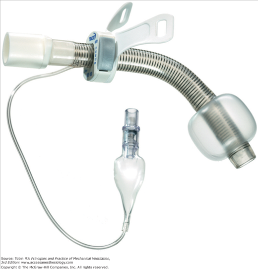
A high index of suspicion for tracheoesophageal fistula is required for critically ill patients who have compatible symptoms that may mimic other swallowing or pulmonary problems. The diagnosis is made by tracheoscopy and esophagoscopy, which determine the location and extent of the fistula. Positioning the cuff of an adjustable tracheotomy tube distal to the fistula (Fig. 40-3) and placement of a gastric tube limit airway soiling by aspirated gastric contents. A jejunostomy tube provides nutritional support. Because spontaneous closure rarely occurs, surgical correction is required, which must be delayed until after weaning from mechanical ventilation to avoid anastomotic dehiscence. Primary closure can correct small tracheal fistulae, but most patients require tracheal resection and reconstruction.276,279 Tracheoesophageal fistulae are universally fatal if left uncorrected.
A tracheoarterial fistula is a major, potentially life-threatening complication of tracheotomy that occurs in less than 1% of patients.280–283 The innominate artery is most commonly involved because it is nine to twelve tracheal rings below the cricoid cartilage within reach of the tip, and sometimes the cuff, of the tracheotomy tube. In some patients, the innominate artery can traverse the midline just below the tracheotomy stoma where the “elbow” of the tube just above the cuff can induce pressure necrosis.284 Arterial erosions, however, may also involve the common carotid artery, inferior thyroid artery, the thyroid ima artery, the innominate vein, and the aortic arch.285 Improper traction on the tracheotomy tube by ventilator tubing, placement of the tracheal window below the fourth tracheal ring, misalignment of the tube with the tip against the tracheal mucosa, high cuff pressures, prolonged intubation, excessive patient movement or posturing, sepsis, malnutrition, and corticosteroid therapy increase the risks for this complication.286–289
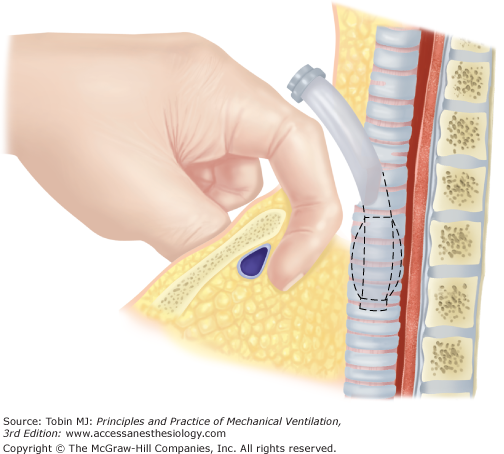
Massive airway hemorrhage and asphyxia are the feared complications of tracheoarterial fistula. Hemorrhage can occur as early as several days or as late as 7 months after tracheotomy with the peak incidence between the first and third weeks.280,283,286,290,291 Up to 50% of patients who experience an episode of airway hemorrhage later than 72 hours after a tracheotomy have an underlying tracheoarterial fistula.286 Pulsations of the tracheotomy tube and “spotty” herald hemorrhages may portend massive hemorrhage. Any airway bleeding 72 hours after tracheotomy warrants consideration of fiberoptic endoscopy in an operating room environment in case airway manipulation precipitates massive hemorrhage.230,280 Three-dimensional CT scanning may establish the diagnosis for stable patients.292 Patients who present with massive hemoptysis should be managed with overinflation of the tracheotomy tube cuff or insertion of a translaryngeal tube with positioning of the cuff over the fistula in an effort to tamponade bleeding. A finger inserted into the stoma tract may allow compression of the innominate artery anteriorly against the sternum (Fig. 40-4).286,287
A tracheoarterial fistula represents a surgical emergency and patients require a median sternotomy with ligation and resection of the artery because tissue infection obviates vascular repair.280,283,285,293 Interruption of the innominate artery is usually tolerated without neurologic sequelae.280,285,293
Epithelialization of the stoma tract may prevent the stoma from closing spontaneously after decannulation. The parastomal platysma or a sternohyoid muscle flap can then be pulled over a surgical stoma repair followed by skin closure.294–296 Some patients develop excessive scarring at a closed stoma site, which can be managed through reconstructive surgery that fills lost deep tissue bulk, corrects any tracheal skin tugging, and produces a tension-free closure that falls more naturally into the neck folds.297
A PDT risks fracture and herniation of tracheal rings because the technique dilates rather than incises intercartilaginous tissue and produces an anteroposterior vector of force at the tracheal insertion site.298–303 Damaged rings can protrude into the airway causing tracheal narrowing304 or promote tracheomalacia if extensive loss of cartilaginous support occurs.305 The frequency of clinically important complications after tracheal ring fractures is unknown. One study followed sixteen patients with tracheal ring fractures and found no instances of tracheal stenosis.306
Timing of Tracheotomy during Mechanical Ventilation
Although translaryngeal endotracheal tubes are relatively well tolerated, patients who require prolonged ventilation eventually benefit from tracheotomy (Table 40-2).307,308 Selecting patients for tracheotomy and identifying the ideal time for the procedure, however, remain clinical challenges with physicians demonstrating considerable practice variation.41,309–311 Clinical trials that randomize patients to “early” versus “late” tracheotomy to inform timing decisions face major difficulties.312 Investigators must be able to identify accurately soon after intubation those patients who will most likely require prolonged ventilation so as to avoid performing unnecessary tracheotomies.313 Unfortunately, experts doubt their own abilities to predict prolonged intubation during the first days of ventilation.314 Recent trials report that a large proportion of enrolled patients randomized to control, nontracheotomy groups based on their high risk of needing prolonged ventilation actually undergo early extubation.315,316 Collected study data should ideally include comprehensive short-term (e.g., pneumonia, incisional bleeding, duration of mechanical ventilation, patient comfort) and long-term (e.g., tracheal stenosis, speech problems, dysphagia) outcomes, which often have nonstandardized definitions, imprecise or costly approaches to detection, and dependency on case mix.
|
Additional study design challenges include (a) physician reluctance to allocate patients to the early tracheotomy group when assigned by randomization, (b) difficulties with blinding providers to group allocation, (c) standardizing patient care with protocols for other critical care interventions and approaches to weaning and extubation, and (d) standardizing the tracheotomy technique (i.e., PDT versus surgical tracheotomy). Consequently, most existing prospective trials have limitations because of small and heterogeneous study populations, variations in defining “early” versus “late” tracheotomy, and various methodological flaws that complicate meta-analyses.
These limitations indicate that timing tracheotomy for critically ill patients remains a complex decision that requires individualization of care. No data exist to establish a standard time when tracheotomy must be performed to avoid airway injury related to continued translaryngeal intubation. Moreover, the risks of performing tracheotomy are acceptably low so that it can be safely applied when patients appear likely to benefit from the procedure. Recommendations to “calendar watch” and avoid tracheotomy if at all possible for at least 14 to 21 days because of its attendant risks have become obsolete. Patients should be selected for tracheotomy by considering the unique circumstances of the patient at hand and the likelihood based on existing studies that the benefits of tracheotomy outweigh its inherent risks as compared with the competing risks of prolonging translaryngeal intubation (Table 40-3). Patient and family values and preferences should also influence the decision.
|
In considering potential benefits, a tracheotomy provides opportunities to improve a patient’s comfort and sense of well-being.316–318 For patients receiving intravenous sedation, some,319,320 but not all,316,321 studies indicate that drug requirements to maintain comfort are decreased after tracheotomy. By freeing the oropharynx, tracheotomy promotes oral hygiene and nutrition and facilitates articulated speech,322–325 allows early mobilization,312,326 and permits transfer from the ICU to more comfortable and family-centered settings for patients who remain ventilator dependent.327 More effective oral care and decreased sedative requirements may decrease risks of ventilator-associated pneumonia.328,329 The largest available randomized trial that assessed impact of tracheotomy timing on pneumonia risk, however, reported no differences between patients undergoing early versus late tracheotomy.315
The shorter length and rigid structure of tracheotomy tubes have been stated to decrease airway resistance and promote weaning from mechanical ventilation.330,331
Stay updated, free articles. Join our Telegram channel

Full access? Get Clinical Tree


