(Reproduced with permission from Shah N, Bedford RF: Invasive and noninvasive blood pressuring monitoring. In: Clinical Monitoring: Practical Applications in Anesthesia and Critical Care Medicine. Lake CL, Hines RL, Blitt CD [editors]. WB Saunders, Philadelphia, 2001, p 182.)
Noninvasive Arterial Blood Pressure Monitoring: Techniques
BP cuffs: Proper cuff size is crucial for an accurate BP measurement. The cuff bladder should extend at least halfway around the extremity, and the cuff width should be 20% to 50% greater than the diameter of the extremity. Incorrect use of automated BP cuffs has resulted in nerve palsies and extravasation of intravenous fluids.
Palpation: SBP (not DBP or MAP) can be determined by occluding flow at a palpable peripheral pulse with a BP cuff. The cuff pressure is released 2 to 3 mm Hg per heartbeat until the pulse is again palpable. SBP is underestimated because of the insensitivity of touch and the delay between pulses and flow under the cuff.
Auscultation: A BP cuff can be inflated to a pressure between SBP and DBP to partially collapse an underlying artery and produce turbulent flow and the characteristic Korotkoff sounds, which are audible to a stethoscope placed under the distal third of the BP cuff. Pressure is measured with a manometer. Korotkoff sounds may be difficult to hear during episodes of hypotension or peripheral vasoconstriction.
Doppler probe: The Doppler Effect is the shift in sound wave frequency when a source moves relative to an observer. A Doppler probe transmits an ultrasonic beam that is reflected by underlying tissue. The probe should be positioned directly above an artery so that the beam passes through the vessel wall. A Doppler probe can be used to detect flow under the BP cuff. Only systolic pressures can be reliably determined with the Doppler technique.
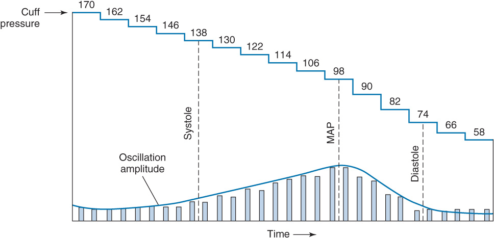
Oscillometry: Arterial pulsations cause small oscillations in cuff pressure when the cuff is inflated above SBP. As cuff pressure decreases closer to SBP, the oscillations increase because of pulse transmission to the entire cuff. Maximal oscillation occurs at MAP, after which oscillations decrease. Automated BP monitors derive SBP, MAP, and DBP after electronically measuring the pressures at which oscillation amplitudes change. Measurements may be unreliable during arrhythmias and when on cardiopulmonary bypass.
Arterial tonometry: Several independent pressure transducers are applied to the skin overlying an artery; beat-to-beat BP is sensed by the pressure required to partially flatten the artery. The contact stress between the transducer directly over the artery and the skin reflects intraluminal pressure. Limitations include movement artifact and the need for frequent calibrations.
Invasive Arterial Blood Pressure Monitoring
Indications: Induced hypotension, anticipated wide blood pressure swings, end-organ disease necessitating precise beat-to-beat blood pressure regulation, and the need for multiple arterial blood samples.
Contraindications: Catheterization should be avoided in arteries of extremities with inadequate collateral blood flow or suspicion of vascular insufficiency (e.g., Raynaud phenomenon).
Selection of Artery for Cannulation
Radial artery: Commonly cannulated because of its superficial location and collateral blood flow. Inadequate collateral flow occurs in 5% of patients because of incomplete palmar arches. Ulnar collateral circulation adequacy can be assessed via the Allen test, palpation, Doppler probe, plethysmography, or pulse oximetry.
Ulnar artery: Deeper and more tortuous than the radial artery. Normally not considered because of a risk of hand blood flow compromise, especially if the ipsilateral radial artery has been punctured.
Brachial artery: Large and easily identifiable in the antecubital fossa and has less waveform distortion because of its proximity to the aorta. Its location predisposes to kinking of the catheter during flexion at the elbow.
Femoral artery: Provides excellent access but is prone to pseudoaneurysm and atheroma formation. The femoral site has been associated with an increased incidence of infections complications and arterial thrombosis, as well as aseptic necrosis of the femoral head in children.
Dorsalis pedis and posterior tibial arteries: The most distorted waveforms because of its distance from the aorta.
Axillary artery: Surrounded by the axillary region of the brachial plexus, and thus nerve damage can result from a hematoma or traumatic cannulation. Flushing of the left axillary artery can easily result in transmission of air or thrombi to the cerebral circulation.
Invasive Arterial Pressure Monitoring: Radial Artery Cannulation Technique
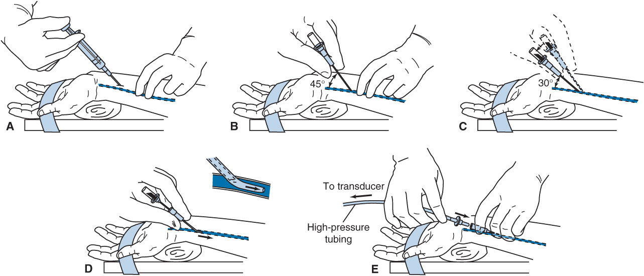
• Supination and wrist extension provide optimal exposure of the radial artery.
• Pressure-tubing-transducer system should be nearby and flushed for easy connection.
• Radial artery course is determined by lightly palpating over the maximal impulse of the radial pulse with the fingertips.
• The skin is prepped with a bactericidal agent, and 0.5 mL of lidocaine is infiltrated directly above the radial artery.
• A 20- or 22-gauge catheter over a needle is passed through the skin at a 45° angle directed toward the point of palpation.
• Upon blood flashback, a guidewire may be advanced through the catheter into the artery and the catheter advanced over the guidewire. Alternatively, the needle is lowered to a 30° angle and advanced 1-2 mm to ensure the catheter tip is in the vessel lumen.
• The needle is withdrawn while firm pressure is applied over the artery proximal to the catheter tip to minimize blood loss as the tubing is being connected.
• Tubing is firmly connected and secured with waterproof tape or suture.
Invasive Arterial Blood Pressure Monitoring: Complications
Complications: Hematoma, bleeding, vasospasm, arterial thrombosis, air embolization, skin necrosis overlying the catheter, nerve damage, infection, necrosis of digits, and unintentional intraarterial drug administration.
Factors associated with increased rate of complications: Prolonged cannulation, hyperlipidemia, repeated insertion attempts, female gender, extracorporeal circulation, and the use of vasopressors.
Complication risk is minimized by the following: When the ratio of catheter to artery size is small, heparinized saline is continuously infused through the catheter at a rate of 2 to 3 mL/h, flushing of the catheter is limited, and meticulous attention is paid to aseptic technique.
Invasive Arterial Blood Pressure Monitoring: Clinical Considerations
Continuous, beat-to-beat measurement via arterial cannulation is the gold standard of BP monitoring.
The transduced waveform depends on the dynamic characteristics of the catheter–tubing–transducer system. Tubing, stopcocks, and air all can lead to overdamping, which will underestimate the systolic pressure.
Underdamping can also occur, which can lead to overshoot and a falsely high, overestimated SBP.
Improve system dynamics: Low-compliance tubing, minimize tubing and stopcocks, remove air bubbles.
Transducers convert the mechanical energy of the arterial pressure wave to an electrical signal, and their accuracy depends on correct calibration and zeroing procedures.
Motion or electrocautery artifacts can result in misleading arterial waveform readings.
The arterial waveform shape provides clues to hemodynamic variables. The rate of upstroke indicates contractility, and the rate of downstroke indicates peripheral vascular resistance. Exaggerated variations in size during the respiratory cycle suggest hypovolemia. MAP is calculated by integrating the area under the pressure curve.
Electrocardiography
Electrodes are placed on the patient’s body to monitor the electrocardiogram (ECG).
The ECG is a recording of myocardial cells’ electrical potentials that allows the detection of arrhythmias, myocardial ischemia, conduction abnormalities, pacemaker malfunction, and electrolyte disturbances.
ECG leads are positioned throughout the body to provide different perspectives of the electrical potentials.
Indications: All patients should have intraoperative ECG monitoring. There are no contraindications.
Electrocardiography: Techniques and Complications
Lead selection determines the diagnostic sensitivity of the ECG.
Lead II—arrhythmias and inferior wall ischemia: Lead II’s electrical axis is ∼60° from the right arm to the left leg, parallel to the atria’s electrical axis, resulting in the largest P wave voltages of any surface lead.
Lead V5—anterior and lateral wall ischemia: Lies at the fifth intercostal space at the anterior axillary line.
A true V5 requires at least five lead wires; a modified V5 can be monitored by three leads.
Electrocardiography: Clinical Considerations
Artifacts are a major problem because of the small voltage potentials being measured. Patient or lead-wire movement, use of electrocautery, 60-cycle interference, and faulty electrodes can simulate arrhythmias.
Depending on equipment availability, a preinduction rhythm strip can be printed or frozen on the monitor’s screen to compare with intraoperative tracings. To interpret ST-segment changes properly, the ECG must be standardized so that a 1-mV signal results in a deflection of 10 mm on a standard strip monitor. Newer units continuously analyze ST segments for early detection of myocardial ischemia.
Central Venous Catheterization
Indications: Central venous pressure (CVP) monitoring, fluid administration to treat hypovolemia and shock, infusion of caustic drugs and total parenteral nutrition, aspiration of air emboli, insertion of transcutaneous pacing leads, and gaining venous access in patients with difficult or poor peripheral veins.
Contraindications: Tumors, clots or tricuspid valve vegetations that could be dislodged during cannulation. Internal jugular vein cannulation is relatively contraindicated in patients who have had an ipsilateral carotid endarterectomy.
Central Venous Catheterization: Techniques and Complications
Placement: A catheter is placed in a vein so that its tip lies at the junction of the superior vena cava and the right atrium. Most central lines are placed using Seldinger technique (catheter over guidewire). The patient is placed in the Trendelenburg position to reduce the air embolism risk and to distend the internal jugular vein. Full aseptic technique must be observed, and ultrasound guidance should be used. It is crucial that the vein is cannulated because carotid artery cannulation can lead to hematoma, stroke, and airway compromise. The risk of vein dilator or catheter placement in the carotid artery can be reduced by transducing the vessel’s pressure waveform or comparing the blood’s PaO2 with an arterial sample. Transesophageal echocardiography can also be used to confirm that the wire is in the right atrium.
Complications: The risks of central venous cannulation include infection, air or thrombus embolism, arrhythmias (indicating that the catheter tip is in the right atrium or ventricle), hematoma, pneumothorax, hemothorax, hydrothorax, chylothorax, cardiac perforation, cardiac tamponade, trauma to nearby nerves and arteries, and thrombosis. Subclavian vein catheterization is associated with significant risk of pneumothorax. Left-sided catheterization carries an increased risk of vascular erosion, pleural effusion, and chylothorax.
Related posts:
 Intravenous Anesthetics
Anesthesia for Patients with Cardiovascular Disease
Anesthesia for Genitourinary Surgery
Ambulatory, Non–Operating Room, and Office-Based Anesthesia
Intravenous Anesthetics
Anesthesia for Patients with Cardiovascular Disease
Anesthesia for Genitourinary Surgery
Ambulatory, Non–Operating Room, and Office-Based Anesthesia
 Spinal, Epidural, and Caudal Blocks
 Cholinesterase Inhibitors and Other Pharmacologic Antagonists to Neuromuscular Blocking Agents
Stay updated, free articles. Join our Telegram channel

Full access? Get Clinical Tree


