(1)
Chennai Breast Centre, Chennai, India
Surgical Handling
Close communication between the surgeon, radiologist, and pathologist is essential for appropriate handling of a breast surgical excision specimen. Good fixation of tissue and high-quality processing and optimal standard H&E staining are prerequisites for optimal pathology reporting.
Well-defined protocols for excision and orientation of surgical specimens should be agreed upon and any deviations from established practice should be noted in the accompanying request form. Specimen orientation can be accomplished by using different lengths of sutures or staples/clips, e.g., long suture-lateral, short superior, medium-medial; (1) clip = anterior, (2) clips = superior, (3) clips = nipple margin. Wire localization specimens should be radiographed, after excision, to confirm the presence and the location of the abnormality. This allows immediate re-excision if the lesion is close to a margin.
The specimen radiographs must also be available to the pathologist to facilitate sampling of the lesion, particularly when the lesion contains only calcifications with no identifiable mass.
The specimen should be immediately placed in an appropriately shaped and sized screw-capped container containing at least twice the volume of 10 % neutral buffered formalin. With larger excisions and mastectomy specimens, the surgeon could make a precise single or cruciate incision over the lesion, taking care not to compromise assessment of margins. This enables rapid fixation, which is essential for good tissue conservation. Poor fixation causes tumor autolysis with consequent deleterious effects on the assessment of mitotic counts and in identifying vascular invasion. Hormone receptor assessment is also hampered by inadequate fixation.
Laboratory Handling
A filled in request form with patient demographics, surgical procedure, details of lesion – side, size, type (calcification, mass, deformity), and other relevant clinical details should accompany the specimen.
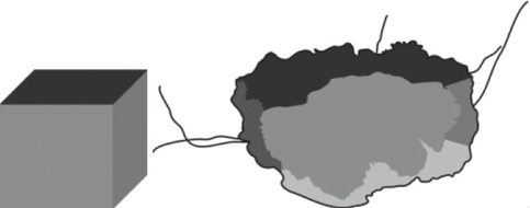
Following receipt in the laboratory, the external surface of the specimen should be painted by using appropriate pigments, such as Indian ink, to enable assessment of margins. Painting can be facilitated by first dipping the specimen in alcohol to clear surface lipid. Fixing of Indian ink can be then be accomplished by using 10 % acetic acid (Fig. 24.1).


Fig. 24.1
Specimen treated like a cube in different colors of Indian ink on each side
Diagnostic Localization Biopsies
The specimen should be weighed, measured in three dimensions, and serially sliced at intervals of approximately 3–5 mm.
Specimens which are approximately 30 mm or less in maximum dimension should be completely embedded and examined histologically.
Larger specimens should be adequately sampled to enable accurate measurement of the size of the lesion.
Excisions performed for mammographically detected lesion such as microcalcification should be serially sliced, laid out in order and x-rayed. Blocks can then be taken from the areas corresponding to the mammographic abnormality, as well as any other suspicious areas identified.
If specimens are sent as more than one piece of tissue, the maximum size of each piece of tissue should be measured and the dimensions added to give an estimated total size.
Therapeutic Wide Local Excision
The approach to sampling a wide local excision depends on the size and type of the specimen. Several methods are described. Any method that is used should enable assessment of important prognostic factors, including greatest lesional dimension and distance to/involvement of margin. The specimen should be weighed and measured in three dimensions.
Method 1: Serial Slicing Perpendicular to the Medial–Lateral or Anteroposterior Plane
Impalpable, screen-detected lesions, particularly microcalcifications, are preferably grossed by this method as the specimen slices can be radiographed to map the position of the calcifications.
After fixation, the specimen is inked to mark excision margins. Using different colored inks on each surface/margin will aid in microscopic identification of specific margins.
The specimen is sliced at intervals of approximately 3–5 mm (in the medial–lateral or anteroposterior plane).
For specimens excised for micocalcifications, the slices should be serially laid out and the specimen re-radiographed. This will enable blocks to be taken from the area of the abnormality. Marking the site of sampling on the specimen radiograph will aid discussions with the radiologist in difficult cases.
The laid out slices should also be photographed. Annotations for the site of sampling (block key) can be marked on the photograph, which can also serve to identify gross distance to excision margins.
Alternately, a detailed diagram can be drawn, marking the margins, the lesion and identifying the site of sampling.
With excisions performed for known DCIS, fibrous breast tissue proximal and distal to the calcification should be sampled, as the DCIS may extend beyond radiologically detected calcifications.
The number of blocks taken will depend on the size of the specimen and the abnormality present. Small specimens (samples less than 30 mm in maximum dimension) can be completely sliced, processed, and examined histologically.
For larger specimens, the area of the mammographic abnormality and the adjacent tissue should be sampled, to avoid underestimation of the maximum dimension of the lesion.
The excision margins of the specimens should also be sampled. The closest margin to the mammographic abnormality must be definitely blocked, to enable microscopic assessment of this distance. As good practice, however, all the margins should be widely sampled to allow a more accurate reading of the completeness of excision.
Specimens containing a palpable or visible macroscopic abnormality are ideally grossed by this method.
A cruciate incision is made over the middle of the tumor, from the posterior/deep fascial plane aspect of the specimen. The tumor is then sampled as four blocks, including the antero–posterior, medial–lateral, and superior–inferior surfaces.
The radial margin and the lesion may be sampled in one block for smaller resections. With large specimens, more than one block may be required to sample the tumor and margin.
Blocks taken through the lesion to the radial edges of the specimen will allow measurement of the lesion to margin distance.
Pathologic shave margin: The circumferential edge of the sample can be shaved to allow more extensive examination of relevant surgical resection margins. A pathologic shaved margin is a thin slice of tissue, usually about 2 mm in thickness, which is taken parallel to the inked surface of the specimen. The tissue is then embedded en face and the margin is regarded as positive if tumor is identified anywhere in the section. The disadvantage of this method is that the tumor may be up to 2 mm away from the true inked margin.
Stay updated, free articles. Join our Telegram channel

Full access? Get Clinical Tree


