![]() Is an intrauterine pregnancy (IUP) (defined as yolk sac or fetal pole) present?
Is an intrauterine pregnancy (IUP) (defined as yolk sac or fetal pole) present?
![]() Abdominal or pelvic pain
Abdominal or pelvic pain
![]() Suspected ectopic pregnancy or risk factors for ectopic pregnancy
Suspected ectopic pregnancy or risk factors for ectopic pregnancy
![]() Vaginal bleeding
Vaginal bleeding
![]() Unexplained syncope, or hypotension
Unexplained syncope, or hypotension
![]() Pelvic mass
Pelvic mass
CONTRAINDICATIONS
![]() Absolute: None
Absolute: None
![]() Relative (transvaginal approach): Recent major pelvic surgery
Relative (transvaginal approach): Recent major pelvic surgery
CONSENT
![]() Get verbal or written consent for the procedure, except in extremis situations
Get verbal or written consent for the procedure, except in extremis situations
RISKS
![]() No documented harmful effects on the fetus or the mother due to ultrasound exposure
No documented harmful effects on the fetus or the mother due to ultrasound exposure
LANDMARKS
![]() Transabdominal
Transabdominal
![]() Have the patient lie supine
Have the patient lie supine
![]() The bladder should be full in order to have an adequate acoustic window
The bladder should be full in order to have an adequate acoustic window
![]() Use a standard curved 3.5- to 5.0-MHz probe to scan the lower abdomen
Use a standard curved 3.5- to 5.0-MHz probe to scan the lower abdomen
![]() Place the probe on the anterior abdominal wall, at the level of the symphysis pubis
Place the probe on the anterior abdominal wall, at the level of the symphysis pubis
![]() For advanced gestations, place the probe more proximally
For advanced gestations, place the probe more proximally
![]() Transvaginal
Transvaginal
![]() Insert the probe into the vaginal canal
Insert the probe into the vaginal canal
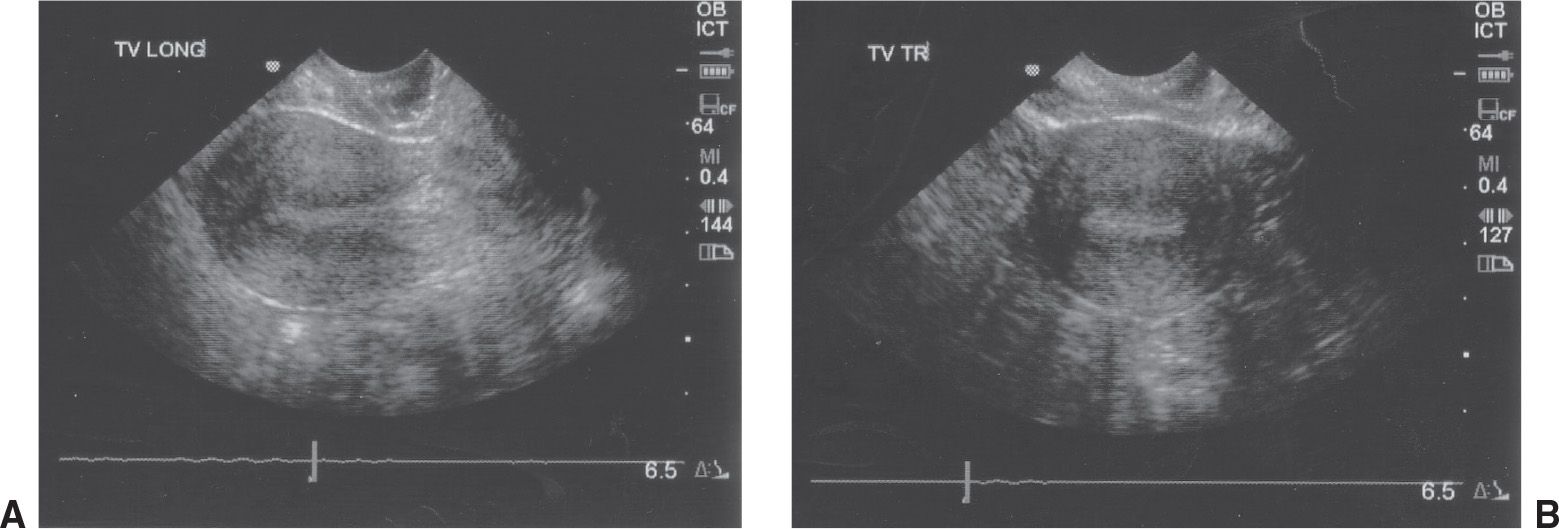
![]() The uterus is midline, posterior to the bladder and anterior to the rectum (FIGURE 44.1)
The uterus is midline, posterior to the bladder and anterior to the rectum (FIGURE 44.1)
![]() The right and left ovaries are lateral to the uterus and anteromedial to the right and left iliac vessels
The right and left ovaries are lateral to the uterus and anteromedial to the right and left iliac vessels
![]() Anteroflexed uterus 90% and retroflexed in 10%
Anteroflexed uterus 90% and retroflexed in 10%
Related posts:
Stay updated, free articles. Join our Telegram channel

Full access? Get Clinical Tree


