ANTIBACTERIAL THERAPY: THE OLD, THE NEW, AND THE FUTURE

CHAPTER 97 ANTIBACTERIAL THERAPY: THE OLD, THE NEW, AND THE FUTURE


Infections remain the leading cause of death in hospitalized patients, and antimicrobial therapy is a mainstay of treatment. However, widespread overuse and misuse of antibiotics have led to an alarming increase in multiple-drug-resistant (MDR) pathogens. New agents may allow shorter courses of therapy and prophylaxis, which are desirable for cost control and control of microbial flora. Moreover, antibiotics are second only to analgesic agents in the number of adverse drug reactions.


PRINCIPLES OF PHARMACOKINETICS


The goal of pharmacotherapy is an effective response with no toxicity. The prescriber must have knowledge of the principles of drug absorption, distribution, and elimination. The dose-response relationship is influenced by dose, dosing interval, and route of administration. The plasma drug concentration is influenced by absorption, distribution, and elimination—which in turn depend on drug metabolism and excretion. The plasma concentration may not reflect tissue concentrations, as penetration into individual tissues is variable. Finally, the relationship between local drug concentration and effect is defined by several pharmacodynamic (PD) principles (see following discussion).


A few basic concepts of pharmacokinetics (PK) are useful to the practitioner. Bioavailability is defined as the percentage of an administered dose of a drug that reaches the systemic circulation. By definition, bioavailability is 100% after intravenous administration. However, this varies among drugs after oral administration, being affected by absorption (a function of product formulation and gastric emptying time), intestinal transit time, and the degree of hepatic first-pass metabolism.


Half-life refers to the amount of time required for the drug concentration to reduce by half, and thus is a hybrid of consider ations of both clearance and volume of distribution. Half-life is useful to estimate when a steady-state drug concentration will be achieved. If a “loading dose” is not administered intravenously, thereby creating instantaneously a desired drug concentration to be maintained throughout therapy, four to five half-lives must elapse to achieve a steady state. Changes in dosage and changes in half-life owing to disease state (e.g., renal failure) must be accounted for. Interpretation of drug concentration data is difficult if the patient is not at a steady state, especially so in critical illness characterized by fluctuating organ function and volume of distribution.


Volume of distribution (VD) is a proportionality constant that relates to plasma concentration and the amount of drug in the body. VD is useful for estimating achievable plasma drug concentrations that result from a given dose. It is a derived parameter that is independent of a drug’s clearance or half-life. It does not have particular physiologic significance, but pathophysiologic conditions can alter VD substantially. A reduction of VD will result in a higher plasma drug concentration for a given dose. However, the “third space” extravascular volume redistribution, fluid overload, and hypoalbuminemia (with decreased drug binding) of surgical illness act to increase VD, all of which makes dosing a complex matter.


Clearance refers to the volume of liquid from which drug is eliminated completely per unit of time (whether by distribution to tissues, metabolism, or elimination) and is important for determining the amount of drug necessary to maintain a steady-state concentration. Drug elimination may be by metabolism, excretion, or dialysis. Most drugs are metabolized by the liver to polar compounds that can then be excreted by the kidney, but metabolism does not imply inactivation. For example, metronidazole is metabolized to a bactericidal metabolite with a prolonged half-life that has dosing implications. The kidneys are most important for excretion of metabolized drugs, although some drugs are metabolized or conjugated by the kidneys. Renal excretion may occur by filtration or by active or passive transport. The degree of filtration is determined by molecular size and charge and by the number of functional nephrons. In general, if greater than 40% of administered drug or its active metabolites is eliminated unchanged in the urine, decreased renal function will require a dosage adjustment. Active reabsorption and concentration of aminoglycosides by proximal tubular cells is a likely component of its well-recognized nephrotoxicity.


PRINCIPLES OF PHARMACODYNAMICS


The variable responses to drugs administered to a heterogeneous patient population can be described and perhaps reduced by an understanding of PD, the relationship of a drug to its intended effect. The PD of antibiotic therapy is especially complex because drugpatient, drug-microbe, and microbe-patient interactions must be accounted for. Knowledge of how patient characteristics influence absorption, distribution, and elimination of a drug—and how an antibiotic interacts with the targeted microbe—can increase the likelihood of a salutary clinical response. In turn, antimicrobial effects on bacteria are highly variable. Microbial physiology, inoculum size, microbial growth phase, intrinsic and extrinsic mechanisms of resistance, microenvironmental factors such as the pH at a local site of infection, and the patient’s immune response are important factors. In the case of antimicrobial therapy, the key drug interaction is not with the host but with the microbe.


Because of microbial ability to alter the nature of the interaction with antimicrobial agents (principally via the development of resistance), mere delivery of drug may not be microbicidal. Factors that may contribute to the development of resistance are the production of drug-inactivating enzymes, alteration of cell surface receptor target molecules, and altered bacterial permeability to antimicrobial penetration. Critical to the microbe-patient interaction is the patient’s immune system. Also inseparable are drug-patient factors that may influence PK, such as hepatic and renal function, serum albumin concentration, and extracellular volume status.


Antibiotic PD is determined by laboratory analysis, and thus the extrapolation of in vitro results to the patient may be challenging because the interaction with the host immune system is isolated from the analysis of the drug-microbe interaction. Analyses from in vitro study include the minimal inhibitory concentration (MIC). The MIC is the minimal serum drug concentration necessary for inhibition of bacterial growth, expressed as the proportion of the inoculum inhibited (MIC90 refers to 90% inhibition). However, some antibiotics may have important effects on bacteria at subinhibitory concentrations. Moreover, MIC testing may not detect the presence of resistant bacterial subpopulations (a particular problem with “heteroresistance” of Gram-positive bacteria, particularly Staphylococcus aureus).


Sophisticated analytic strategies draw upon the principles of both PK and PD; for example, by determination of the peak serum concentration:MIC ratio, the duration of time plasma concentration remains above the MIC, and the area of the plasma concentration-time curve above the MIC (the “area under the curve,” or AUC). With some agents, antibacterial effects may persist for prolonged periods after the plasma drug concentration has become “subtherapeutic.” The persistent inhibition of bacterial growth (but not killing) that persists after the serum drug concentration has fallen below the MIC for the organism is known as the postantibiotic effect (PAE). Appreciable PAE can be observed with amino-glycosides and fluoroquinolones for Gram-negative bacteria, and with some β-lactam drugs (notably carbapenems) against S. aureus. Through analyses of this type, certain drugs (e.g., aminoglycosides) have been characterized as having concentration-dependent killing whereby a higher peak concentration increases the efficacy of bacterial killing (up to a point). Other agents (most β-lactam agents) exhibit bactericidal properties that are independent of concentration. Rather, efficacy is determined by the duration of time the plasma concentration remains above the MIC. Other agents (e.g., fluoroquinolones) exhibit both properties such that bacterial killing may increase as drug concentration increases up to a point of saturation, after which the effect becomes independent of concentration.


EMPIRIC ANTIBIOTIC THERAPY


The decision to administer empiric antibiotic therapy must be considered carefully. An injudicious approach could result in nontreatment of established infection or therapy when the patient has only sterile inflammation or colonization with bacteria. Inappropriate therapy (e.g., delay, therapy misdirected against usual pathogens, failure to treat MDR pathogens) leads unequivocally to increased mortality. Several questions should be asked in each circumstance where empiric therapy is being considered.


Are antibiotics indicated at all? The answer is ultimately often no, but the decision to start treatment of the unstable patient must often be made before definitive information becomes available. The decision to start antibiotics empirically is based on the likelihood of infection, its likely source, and whether the patient’s condition is sufficiently precarious that a delay will be detrimental. Outcome from serious infections is improved if antibiotics are started promptly, but on the other hand only about 50% of fever episodes in hospitalized patients are caused by infection. Many causes of the systemic inflammatory response syndrome are not due to infection (e.g., aspiration pneumonitis, burns, trauma, pancreatitis), although they may be complicated later by infection. Multiple organ dysfunction syndrome may progress even after an infectious precipitant has been controlled, due to a dysregulated host response.


Must antibiotics be started immediately? If the presumed infection is not destabilizing, this decision also depends on the overall status of the patient and should take into consideration such host factors as age, debility, renal and hepatic function, and immunosuppression. Culture yields are highest before antibiotics are administered, which for certain types of specimens (e.g., blood, cerebrospinal fluid) can be crucial. However, for many infections (e.g., bacteremia, intraabdominal infection, pneumonia) early appropriate therapy improves outcome.


Which organisms are the likely pathogens, and are they likely to be MDR? The clinical setting must be considered (e.g., nosocomial versus community-acquired infection, recent antimicrobial therapy), as must the patient’s environment (e.g., recent hospitalization, proximity to another infected patient, the presence of MDR pathogens in the unit) and any recent microbial cultures obtained from the patient.


Will a single antibiotic suffice? The likely diagnosis and the nature of the probable pathogens are crucial determinants. If a nosocomial Gram-positive pathogen is suspected (e.g., wound or surgical site infection, catheter-related infection, prosthetic device infection, pneumonia) and methicillin-resistant S. aureus (MRSA) is endemic, empiric vancomycin (or linezolid) is appropriate. Some authorities recommend dual-agent therapy for serious Pseudomonas infections (i.e., an antipseudomonal β-lactam drug plus an aminoglycoside). It is important to use at least two antibiotics for empiric therapy of any infection that may be caused by a Gram-positive or Gram-negative infection (e.g., nosocomial pneumonia).


Duration of Therapy


Perhaps the most difficult issue is identifying the endpoint. If bona fide evidence of infection is evident, treatment is continued as indicated clinically. Often, however, the cultures will return negative and the decision must be arbitrary. The decision is complicated further when the patient has had a clinical response to antibiotic therapy in the absence of corroborating evidence, which may be coincident with or a result of false-negative cultures. Moreover, the bias to do something to treat the patient (i.e., continue antibiotic therapy) can be compelling in a patient who is deteriorating.


It must be recognized that careful culture techniques and specimen handling, combined with current sophisticated microbiology laboratory support, make it unlikely that substantive pathogens will be missed. Therefore, continuing empiric antibiotic therapy beyond 48 hours becomes difficult to justify. There are two possible exceptions. One occurs when fungal infection is suspected because the organisms can be difficult to culture, and the other occurs when deep cultures are needed from areas that are inaccessible without radiologic-guided aspiration and some time is necessary to make appropriate arrangements (but is not an excuse for procrastination).


How long should a course of therapy be continued? Effective broad-spectrum antibiotics are widely available, and many infections can be treated with therapy lasting 5 days or fewer. It is important that every decision to start antibiotics must be accompanied by a decision regarding the duration of therapy. A reason to continue therapy beyond the predetermined endpoint must be compelling. Bacterial killing is rapid in response to effective agents, but the host response may not subside immediately. Therefore, the clinical response of the patient should not be the sole determinant for continuation of therapy. If a patient still has sepsis syndrome at the end of a defined course of therapy, it is more useful to stop therapy and obtain a new set of cultures to look for new sites of infection, resistant pathogens, and noninfectious causes of inflammation.


There is a clear trend toward shorter courses of antibiotics for established infections. Broad-spectrum antibiotics that achieve excellent tissue penetration have been an important clinical development, but they also carry morbidity. The worldwide emergence of MDR Gram-positive and Gram-negative bacteria, superinfections in immunosuppressed patients, and the increased mortality associated with nosocomial infections in general make it important that adequate therapy be provided rapidly and for the shortest possible duration. Unfortunately, duration of therapy is not well established in the literature—and new studies are seldom designed with duration of therapy as a primary endpoint. Much depends on expertise and clinical judgment, which is accumulating in favor of shorter courses of therapy. Nowhere is this clearer than for peritonitis and intra-abdominal abscess, for which the previous standard 7- to 10-day courses of therapy have been reduced to 5 days.


Infections that require 24 hours of therapy or less (sometimes just a single dose) include uncomplicated acute appendicitis or cholecystitis, uncomplicated bacterial cystitis (with some agents), and intestinal infarction without perforation. There is seldom justification to continue antibacterial therapy for more than 10 days. Examples of bacterial infections that require more than 14 days of therapy include tuberculosis of any site, endocarditis, osteomyelitis, and selected cases of brain abscess, liver abscess, lung abscess, some cases of postoperative meningitis, and some cases of endophthalmitis. Among the many reasons to limit therapy to only that which is needed is that antibiotic therapy has adverse consequences, despite a widespread perception that therapy is safe if not entirely benign. Adverse consequences of antibiotics include allergic reactions; development of nosocomial superinfections, including fungal infections, enterococcal infections, and Clostridium difficile–related disease; organ toxicity; promotion of antibiotic resistance; reduced yield from subsequent cultures; and induced vitamin K deficiency with coagulopathy or accentuation of warfarin effect.


CHOICE OF ANTIBIOTIC


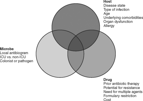
The choice of which antibiotic to prescribe is made based on several interrelated factors. Paramount is activity against identified pathogens, presuming that a distinction between infecting and colonizing organisms can be made and that narrow-spectrum coverage is always most desirable. Knowledge of antimicrobial resistance patterns, nationally and especially in one’s own institution and unit, is essential. Also important is an assumption regarding likely pathogens, which is paramount in cases where empiric therapy is necessary. Estimation of likely pathogens depends on the disease process believed responsible, whether the infection is community- or hospital-acquired, whether MDR organisms are present, and proximity to other infected patients. Also important are patient-specific factors, including age, debility, immunosuppression, intrinsic organ function, prior allergy or another adverse reaction, and recent antibiotic therapy. Institutional factors that may play a role include the existence of guidelines or practice parameters that may specify a particular therapy, or the availability of specific agents as defined by inclusion on the formulary or restriction by antibiotic control programs (Figure 1).


Development of Bacterial Resistance


In general, bacteria use four different mechanisms to develop resistance to antibiotics. Cell wall permeability to antibiotics is decreased by changes in porin channels (especially important for Gram-negative bacteria with complex cell walls, affecting aminoglycosides, β-lactam drugs, chloramphenicol, sulfonamides, tetracyclines, and possibly quinolones). Production of specific antibiotic-inactivating enzymes by plasmid-mediated or chromosomally mediated mechanisms affects aminoglycosides, β-lactam drugs, chloramphenicol, and macrolides. Alteration of the target for antibiotic binding in the cell wall affects β-lactam drugs and vancomycin, whereas alteration of target enzymes can inhibit β-lactam drugs, sulfonamides, quinolones, and rifampin. Drugs that bind to the bacterial ribosome (aminoglycosides, chloramphenicol, macrolides, lincosamides, streptogramins, and tetracyclines) are also susceptible to alteration of the receptor on the ribosome. Antibiotics may be extruded actively once entry to the cell is achieved in the case of macrolides, lincosamides, streptogramins, quinolones, oxazolidinones, and tetracyclines.


Cephalosporin resistance among Gram-negative bacilli can be the result of induction of chromosomal β-lactamases after exposure to the antibiotic. The extended-spectrum cephalosporins are rendered ineffective when bacteria such as enteric Gram-negative bacilli mutate to constitutively produce a β-lactamase that is normally an inducible enzyme. Although resistance to cephalosporins can occur by several mechanisms, the appearance of chromosomally mediated β-lactamases has been identified as a consequence of the use of third-generation cephalosporins. Resistance rates decline when use is restricted. The induction of an extended-spectrum β-lactamase (ESBL) in Klebsiella by ceftazidime was first reported approximately 20 years ago, but more than 200 mutations have now been described in several species of Gram-negative bacteria. The mutant bacteria develop resistance rapidly not only to all cephalosporins but to entire other classes of β-lactam antibiotics. It is therefore justifiable to restrict the use of ceftazidime, especially in institutions grappling with an ESBL-producing bacterium. The carbapenems generally retain useful microbicidal activity against ESBL-producing strains. Increasingly, Pseudomonas aeruginosa produces beta-lactamases of the ampC type.


Quinolone resistance, which is increasing rapidly, is for the most part chromosomally mediated, primarily by changes in the target sites for the antibiotic (DNA gyrase or topoisomerase IV). Changes in permeability or efflux may sometimes cause resistance to quinolones as well. Quinolone resistance is relatively easy to induce if a less-than-maximally effective drug is chosen for initial therapy. Resistance to one quinolone may also increase the MIC for the other quinolones against the organism, and thus if a quinolone is used, a highly active agent given in adequate dosage is essential.


ANTIBIOTIC SPECTRUM OF ACTIVITY


Susceptibility testing of specific organisms is necessary for management of serious infections (including all nosocomial infections). Recommended agents for specific organisms are guidelines only because in vitro susceptibilities may not correlate with clinical efficacy. The necessary concentration of antibiotics may not be achieved in tissue because of underdosing or poor tissue penetration. Moreover, bacterial killing correlates well with peak serum antibiotic concentrations for some drugs (e.g., aminoglycosides) and disorders (e.g., bacterial endocarditis) but correlates better with the duration of bactericidal drug concentrations for other antibiotics (e.g., β-lactam agents).


Penicillins


With the exception of carboxy- and ureidopenicillins, penicillins do not retain important activity against most strains of Gram-negative bacilli. Penicillin G (parenteral) and V (oral) are useful against most strains of aerobic and anaerobic streptococci (except for the increasingly important problem of penicillin-resistant pneumococci [PRSP, up to 40% of isolates] in bacteremia, recurrent otitis, and upper respiratory tract infections). Penicillins also have activity against Enterococcus faecalis (but not E. faecium), Corynebacterium diphtheriae, and Listeria monocytogenes. Gram-negative bacteria that are susceptible to penicillins include Neisseria meningitidis (highly resistant strains exist), some strains of Proteus mirabilis, and Pasturella multocida. In addition to anaerobic streptococci, penicillins are effective against other anaerobes, such as Bacteroides melaninogenicus (but not B. fragilis) and all clostridial species other than C. difficile.


The penicillinase-resistant semisynthetic penicillins include methicillin, nafcillin, oxacillin, cloxacillin, and dicloxacillin. Although these agents have useful activity against streptococci, C. diphtheriae, and anaerobic streptococci, the primary use of these agents is as therapy for sensitive strains of staphylococci. Hospitalized patients who need empiric therapy should not be treated with these agents because 60% of strains of S. aureus (MRSA), 90% of strains of S. epidermidis (MRSE), and virtually all enterococcal strains are resistant. However, these drugs are the treatment of choice for infections caused by susceptible isolates of S. aureus.


Activity against Gram-negative organisms was achieved initially by the addition of an amino group to the penicillin nucleus, thereby creating such drugs as ampicillin and amoxicillin. These drugs retain their antistreptococcal activity and a similar spectrum against most other Gram-positive pathogens, including anaerobic streptococci, but do not have appreciable activity against staphylococci. Ampicillin is highly effective against E. faecalis, including some vancomycinresistant strains (VRE), but only rarely effective against E. faecium. Useful activity remains against N. meningitidis, Moraxella catarrhalis, community-acquired strains of E. coli and Klebsiella spp., Salmonella and Shigella spp., and Proteus spp. Ampicillin remains reasonably effective against community-acquired strains of Hemophilus influenzae, but H. influenzae is increasingly important as a nosocomial pathogen and resistant strains are recognized.


The carboxypenicillins (ticarcillin and carbenicillin) and ureidopenicillins (azlocillin, mezlocillin, and piperacillin; sometimes referred to as acylampicillins) have enhanced activity against Gram-negative bacteria and some activity against P. aeruginosa. Ureidopenicillins have greater intrinsic activity against Pseudomonas, but with the advent of β-lactamase inhibitor combination drugs none of these agents is used widely anymore. Beta-lactamase inhibitors (sulbactam, tazobactam, and clavulanic acid) result in enzymatic inactivation and enhanced effectiveness of the antibacterial agent. The effectiveness of these drugs as antibacterial agents is primarily a function of the inherent antibacterial properties of the parent compound (ampicillin < ticarcillin < piperacillin), and to a lesser extent of the effectiveness of the inhibitor (sulbactam ∼ clavulanic acid < tazobactam). The spectrum of activity varies as a result, and the treating clinician needs to be familiar with each of the drugs in this class.


All of these drugs are effective against streptococci, methicillin-sensitive strains of S. aureus, Listeria monocytogenes, Salmonella, Proteus, and Providentia spp., P. multocida, and widely effective against anaerobes—including anaerobic cocci, B. fragilis, Bacteroides and Prevotella spp., and Clostridium spp. (except for C. difficile). Piperacillin/tazobactam has the widest spectrum of activity against Gram-negative bacteria, and the most potency against P. aeruginosa. Although ampicillin/sulbactam has excellent activity against community-acquired Gram-negative bacilli, it has major shortcomings against hospital-acquired strains of E. coli and Klebsiella (as many as 50% of strains may be resistant). However, sulbactam has useful activity against Acinetobacter spp., making ampicillin/sulbactam an option for therapy of infections caused by susceptible strains.


Cephalosporins


More than 20 antibiotics comprise this class of agents. The characteristics of the drugs thus vary widely when considered individually. It is useful to consider these drugs within four broad “generations” whose general characteristics are similar. For example, the firstgeneration agents retain useful activity against Gram-positive organisms—whereas the second-generation agents generally lose that activity in favor of antianaerobic activity. In contrast, the thirdgeneration agents generally have enhanced activity against Gram-negative bacilli—and some have specific antipseudomonal activity. However, most lack efficacy against Gram-positive organisms and none is effective against anaerobic bacteria.


Cefepime, the fourth-generation cephalosporin available in the United States, has enhanced antipseudomonal activity and has regained activity against most Gram-positive cocci but not MRSA. None of the cephalosporins, regardless of class, has clinically useful activity against any of the enterococci. Regardless, there is sufficient heterogeneity of spectrum (especially among the third-generation agents) such that the clinician should be familiar with all of these drugs. Collectively, they account for a majority of prescriptions for parenteral antibiotics. Ceftriaxone, a third-generation agent unique in its class for excellent activity against Gram-positive organisms and once-daily dosing, was at one time the most-prescribed injectable antibiotic worldwide.


Third-Generation Cephalosporins


Rightly or wrongly, third-generation cephalosporins dominate prescribing practices for parenteral antibiotics. These agents include cefoperazone, cefotaxime, cefpodoxime, cefprozil, ceftazidime, ceftibuten, ceftizoxime, ceftriaxone, and lorcarbicef. They are relatively resistant to β-lactamases, and therefore have an extended spectrum of activity against Gram-negative bacilli. Despite this, these agents lack efficacy against Gram-positive bacteria (except for ceftriaxone) and anaerobic bacteria. Activity is reliable against non-ESBL–producing species of Enterbacteriaceae, including Enterobacter, Citrobacter, Providencia, and Morganella. Activity is variable against Acinetobacter and the pseudomonads, with broad activity against Aeromonas, reasonable albeit variable activity against P. aeruginosa (cefoperazone and ceftazidime), but no activity against S. maltophilia. Ceftriaxone and ceftazidime have activity against Borrelia burgdorferi, the agent of Lyme disease.


Paradoxically, third-generation cephalosporins (particularly ceftazidime) have been associated with the induction of ESBLs among many of the Enterobacteriaceae. Production of ESBLs was first reported in strains of Klebsiella pneumoniae, but now is so well recognized that susceptible pathogens are now referred to commonly as “inducible enteric” bacteria. The resistance induced by ESBL production is not just against other third-generation cephalosporins but affects entire other classes of β-lactam antibiotics. Third-generation cephalosporins, especially ceftazidime, have also been implicated (in concert with the widespread overuse of vancomycin; see material following) in the emergence of VRE. Because resistance can be transferred between enterococci and staphylococci, staphylococci of intermediate susceptibility to glycopeptides (GISA) or resistant to vancomycin (VRSA) have now been reported. Because of the potential to induce resistance of hospital flora, many centers no longer use third-generation cephalosporins as empiric therapy but rather reserve them for directed narrow-spectrum monotherapy of known susceptible organisms.


< div class='tao-gold-member'>

Stay updated, free articles. Join our Telegram channel

Jul 7, 2016 | Posted by in CRITICAL CARE | Comments Off on ANTIBACTERIAL THERAPY: THE OLD, THE NEW, AND THE FUTURE

Full access? Get Clinical Tree

Get Clinical Tree app for offline access