ADVANCED TECHNIQUES IN MECHANICAL VENTILATION

CHAPTER 87 ADVANCED TECHNIQUES IN MECHANICAL VENTILATION


Since the introduction of mechanical ventilation using a bicycle tire and bellows about 50 years ago, the science and art of respiratory therapy has advanced dramatically—allowing the clinician to ventilate and oxygenate patients who would have died in the past due to limitations of man and machine. This chapter focuses on recent advances and future considerations in ventilatory support to allow further improvements in respiratory care and survival from acute lung injury (ALI), acute respiratory distress syndrome (ARDS), and chronic respiratory failure.


IMPROVING OXYGENATION AND PREVENTING ACUTE LUNG INJURY


Providing adequate oxygen delivery (DO2) with minimal barotrauma is the primary goal of mechanical ventilation for patients with all types of pulmonary pathology, as well as for those with normal lungs. Noninvasive ventilation (NIV) using such modalities as bi-level positive airway pressure (BiPAP) with various degrees of inspiratory and expiratory pressure applied via a face, nasal, or combined face mask has become a more common modality to avoid endotracheal intubation or to perhaps shorten the need or period of ventilation by artificial airway. Marginal candidates for liberation from ventilation may stave off reintubation once extubated.


Peak and plateau airway pressures are crucial parameters for the clinician in managing patients on the ventilator. The ARDSnet trial examined conventional mechanical ventilation with a tidal volume (VT) of 10 ml/kg and with VT at a lower tidal volume of 6 ml/kg in patients with ALI/ARDS and found a significant improvement in oxygenation, a decrease in ventilator-associated lung injury (VILI), and decreased mortality related to ALI/ARDS. Tidal volumes as low as 4 ml/kg may be used to maintain the plateau pressure at less than 30 cm H2O to minimize barotrauma (or “volutrauma,” as it is called by some). In some circumstances, conventional mechanical ventilator modalities may be inadequate to the task. Modern microprocessor-controlled ventilators allow modification of flow rate and flow patterns in providing adequate and safe mechanical ventilation.


Ventilator-Associated Lung Injury


Acute lung injury and ARDS are recognized as affecting the lungs heterogeneously. The distribution of edema fluid, ventilated versus flooded alveoli, and consequently the matching of ventilation and perfusion vary among gas exchange units. Moreover, it is recognized that the lung is capable of a brisk inflammatory response when injured or when ventilated mechanically, which may have local or systemic manifestations. The ARDSnet trial demonstrated improved outcomes from ALI/ARDS after ventilation with lower VT and minute ventilation (VE), resulting in lower airway pressures, less overdistension of recruitable alveoli, less shear stress on lung tissue, and lower mortality despite the paradox that most patients with ALI/ARDS do not die from an inability to oxygenate or ventilate. Rather, most such patients die in association with the multiple organ dysfunction syndrome—which has been linked closely with a rampant systemic inflammatory response. If less ventilation is better, it was hypothesized that more ventilation may be injurious or indeed provocative to the lung—leading to the concept of VILI.


Ventilator-induced lung injury occurs from excessive mechanical stress to the lung, either from excessive VT or excessive airway pressure. Mechanical ventilation induces a pulmonary and systemic cytokine response, which can be minimized by limiting overdistension and phasic recruitment/derecruitment of lung. A substantial body of experimental and clinical data demonstrates that the mechanism of VILI is the proinflammatory response in the lung and the periphery, and that the response and injury are attenuated by lung-protective ventilation strategies. New modes of ventilation and protective ventilation are designed to minimize the deleterious effects of mechanical ventilation, which is a fundamental aspect of critical care management.


ALTERNATIVES TO CONVENTIONAL MECHANICAL VENTILATION


Proportional Assist Ventilation


Proportional assist ventilation (PAV) is a form of synchronized partial ventilatory assistance that augments the flow of gas to the patient in response to patient-generated effort. The ventilator augments the patient’s inspiratory effort without using preselected target volume or pressure. The purpose of PAV is to allow the patient to achieve a pattern of ventilation and breathing that is adequate and comfortable. The patient initiates and determines the depth and frequency of the breaths independently of the ventilator. Advantages to this type of ventilator support include greater comfort; reduction of peak airway pressure required to deliver the VT; less likelihood of overventilation and overdistension of alveoli; preservation and enhancement of the patient’s own reflex, behavioral, and homeostatic control mechanisms; and improved efficiency of negative-pressure ventilation.


Effective use of PAV requires an understanding of the individual patient’s ventilatory mechanics. This entails measuring the patient’s airway resistance, compliance, and intrinsic positive end-expiratory pressure (auto-PEEP) to determine the ventilatory load and assistance the patient requires. Younes et al. proposed an innovative method for the noninvasive determination of passive elasticity during PAV. Once the patient’s elastance and resistance are determined, the PAV parameters are set followed by PEEP, adjusting the peak pressure limit to 30 cm H2O, adjusting volume assist to 8% of elastance measured on PAV, and finally observing the patient’s ventilation, breathing pattern, and peak airway pressure. As a new ventilatory method, PAV can conceivably improve patient-ventilator interaction. Its true usefulness remains to be measured, and clinical usage is uncommon.


Mandatory Minute Ventilation


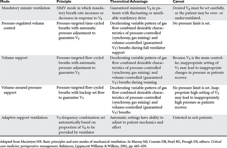
Mandatory minute ventilation (MMV) is a mode of mechanical ventilation in which the minimum level of VE needed by the patient is provided. If the patient’s spontaneous ventilation is insufficient to meet the predetermined VE, the ventilator provides the difference. Conversely, if the patient’s spontaneous breathing exceeds the target VE no ventilator support is provided. This mode is one of the so-called “closed-loop” ventilation modes (Table 1) because the ventilator varies its parameters in response to the patient’s own intrinsic ventilatory requirements. The major advantage of MMV is the capability to vary ventilatory support according to the response of the patient. This mode of mechanical ventilation is best suited for patients with severe neuromuscular disease or drug overdose, or patients heavily sedated. One of the main disadvantages with MMV is that alveolar ventilation may not be matched equally with exhaled VE, thus diminishing closing volumes and leading to atelectasis. None of the closed-loop modes, MMV included, has been tested sufficiently on critically ill patients to recommend widespread incorporation into practice.


Airway Pressure Release Ventilation


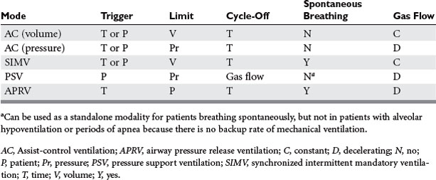
Airway pressure release ventilation (APRV) (Table 2) has been used as an alternative mode of mechanical ventilation in patients with acute respiratory failure. APRV, which has been available in some ventilator models since the mid-1990s, allows for the unloading during exhalation of any positive pressure provided during inhalation in order to facilitate the egress of the tidal breath. Release of airway pressure from an elevated baseline simulates exhalation. Technically, APRV is time-triggered, pressure-limited, time-cycled mechanical ventilation. Conceptualizing APRV as continuous positive airway pressure (CPAP) with regular, brief, intermittent releases of airway pressure may facilitate understanding. It can augment alveolar ventilation in the patient breathing spontaneously, or provide full support to the apneic patient.


Advantages of APRV include lower peak airway pressure, lower intrathoracic pressure, lower VE, minimal effect on cardiac output, and improved matching of ventilation and perfusion. The mode may facilitate spontaneous breathing by the patient. Sedation requirements may be decreased, and neuromuscular blockade should be avoided altogether. Patient-ventilator dyssynchrony is believed not to develop. Disadvantages of APRV include pressure control of ventilation, increased effects of airway and circuit resistance on ventilation, decreased transpulmonary pressure, and potential interference with spontaneous ventilation. Facilitated exhalation may make APRV beneficial in patients with bronchospasm or small-airways disease. This mode of ventilation can be used as a weaning mode. Although increasingly popular, the advantage of APRV over other modes of ventilation is unproved.


The terminology of APRV differs somewhat from other modes of mechanical ventilation, and has yet to be standardized. Four important terms include pressure high (Phigh), pressure low (Plow), time high (Thigh), and time low (Tlow). The Phigh term describes the baseline airway pressure (the higher of the two pressures), alternatively called CPAP, inflating pressure, or the P1 pressure. The Plow term describes the airway pressure resulting from the release of pressure (alternatively called PEEP, release pressure, or the P2 pressure). The Thigh time refers to the time during which Phigh is maintained (T1), whereas Tlow refers to the duration of time when airway pressure is released (T2). Mean airway pressure can be calculated from the following equation:


image


Application of APRV to the patient must be individualized, as standard approaches have yet to emerge. Initial settings are deduced partly from the result of conventional mechanical ventilation, which should be attempted initially for most patients. The plateau airway pressure (Pplat) from conventional ventilation (if not higher than 35 cm H2O) is converted to Phigh, aiming for a VE of 2–3 l/min (lower than with conventional ventilation). The Plow pressure is set initially at 0 cm H2O. The setting for Thigh is a minimum of 4 seconds, and Tlow is set at approximately 0.8 seconds (0.5–1.0 second). Spontaneous breating is permitted. At these settings, mean airway pressure is 29 cm H2O. Rarely, a higher Phigh (40–45 cm H2O) is needed for patients with low compliance (e.g., morbid obesity, abdominal distention). For all patients, Thigh is lengthened progressively to 12–15 seconds (usually in 1- to 2-second increments as lung mechanics improve). Longer Thigh prevents the cyclical opening and closing of small airways that is believed to be a cause of VILI. The Tlow parameter is optimized when expiratory flow decreases to 25%–50% of peak expiratory flow.


Clinical improvement may not be immediate after transition to APRV (as is the case with IRV). Clinical studies have shown that maximum clinical improvement may not occur until 8–16 hours after the transition. After improvement, weaning from APRV is guided by general principles of weaning. Weaning from APRV is accomplished primarily by manipulation of Phigh and Thigh. High pressure is decreased in increments of 2–3 cm H2O down to about 15 cm H2O, and Thigh is lengthened progressively to 12–15 seconds (usually in 1- to 2-second increments). Minute ventilation must be monitored carefully for signs of hypoventilation during the transition. The goal is to switch the patient to pure CPAP of 6–12 cm H2O, at which point the patient may be extubated—all conditions permitting.


Some confusion arises with similar modes of ventilation. BiPAP differs from APRV only in the timing of Thigh and Tlow. The latter is longer in BiPAP. Intermittent mandatory pressure release ventilation (IMPRV)—similar to APRV and rarely used—synchronizes the release of pressure with the patient’s spontaneous effort. In IMPRV, all spontaneous breaths are pressure-supported ventilation (PSV) to reduce the work of breathing. However, the rationale for IMPRV is considered dubious by some because dysynchrony appears not to occur with APRV.

< div class='tao-gold-member'>

Stay updated, free articles. Join our Telegram channel

Jul 7, 2016 | Posted by in CRITICAL CARE | Comments Off on ADVANCED TECHNIQUES IN MECHANICAL VENTILATION

Full access? Get Clinical Tree

Get Clinical Tree app for offline access