Acute and chronic pain in the thoracic spine plays only a minor role compared to that of the cervical and lumbar spine. This applies both to the incidence and to the severity of symptoms: Only 2% of all painful spinal syndromes affect the thoracic spine. In very rare cases, therapy-resistant nerve root irritation (intercostal neuralgia) occurs that requires surgical treatment.
Specialized Thoracic Neuroanatomy
The spinal canal is relatively narrow in the thoracic area and there is only a slender epidural space between the spinal cord, the bony surroundings, and the intervertebral disk. The canal is at its narrowest between T4 and T9.
The thoracic spine consists of the zygapophyseal joints, as well as the joints between the vertebrae and ribs (costovertebral joints, costotransverse joints), which jut out from the inferior part of the intervertebral foramen and push the spinal nerve out into the open section above (Fig. 8.1).
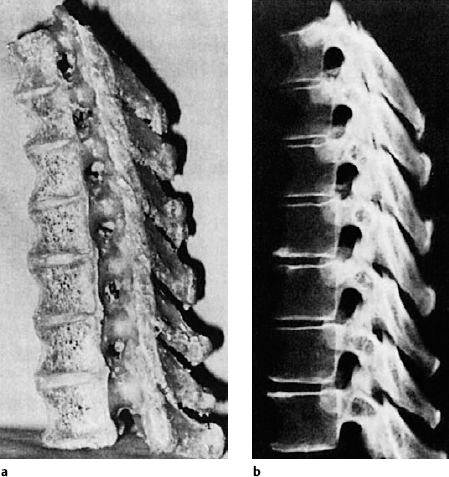
Because of the relatively large diameter of the intervertebral foramen, the osteogenic constrictions that are commonly found in the cervical spine, for example, are rarely seen in the thoracic spine. The intervertebral foramen is not adjacent to the intervertebral disk, as is the case in the cervical and lumbar spine. Rather, it is located at the same level as the vertebral body (Fig. 8.2 a, b).
The displacement of the spinal cord segments in relation to the corresponding vertebral motor segments, as previously described for the cervical spine, continues in the thoracic spine. Between the 1st and 6th thoracic spinous process the displacement amounts to the height of two segments; between the 7th to 10th spinous processes, three segments. Ventral branches of the thoracic spinal nerve, the intercostal nerve, supply the wall of the rib cage, i. e., the intercostal muscles, the costotransverse joints, the parietal pleura, and the skin. A so-called intercostal neuralgia evolves when the thoracic spinal nerves are irritated.
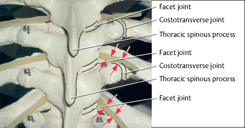
Fig. 8.1 Segments of the mid-thoracic spine on a skeleton, posterior view. In addition to the thoracic facet joints, the thoracic spine also contains costotransverse joints that jut out from the lower part of the intervertebral foramen and push the spinal nerve (arrows) into the freely open section above.
Clinical Picture
The symptoms arising from a compromised thoracic spinal nerve root are characterized by a girdle-shaped pattern of pain with eventual discrete algesic disorders. Its topology is based on the dermatomal pattern. The boundaries between the individual dermatomes are not as well defined in the thoracic area as they are in the peripheral sections of the limbs. Another important diagnostic criterion for intercostal neuralgia due to degenerative changes is the dependency on position of the thoracic spine. Pain is relieved when the thoracic spine is unloaded or extended. Pain increases on loading and with certain rotary movements of the body. This provides information about how the condition should be treated.
Unloading the thoracic vertebral motor segments using the horizontal position is important in therapy. The application of all types of heat treatment is perceived as pleasant because the heat relieves the reflex hypertonicity of the trunk muscles, in particularly the paravertebral back extensors, and stimulates blood flow. These techniques are complemented by the use of manipulation, mainly in the form of traction, physiotherapy exercises to strengthen the trunk muscles, and local injections.
The irritation of thoracic spine nociceptive afferents is considerably more common than the irritation of spinal nerves. This form of irritation arises from incorrect posture or loading, or in some cases, segmental dysfunction or degenerative changes. It can cause extremely painful and treatment-refractory reflexive pain syndromes in the thoracic spine.
So-called viscerovertebral pain syndromes and pain syndromes associated with psychosocial problems can also manifest themselves in the thoracic spine.
Thoracic Injection Therapy
Thoracic Spinal Nerve Analgesia
Principle
Posterolateral injection of a local anesthetic (mixed with steroids when necessary) into the foraminoarticular region of the vertebral motor segment.
Indication
Any therapy-resistant intercostal neuralgia or intercostal neuralgia-like pain with girdle-shaped ipsilateral or bilateral radiation is an indication for thoracic spinal nerve analgesia (TSPA) treatment.
Technique
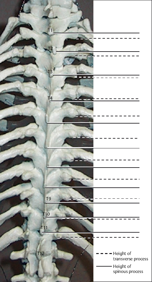
In order to carry out TSPA, bony contact has to be made with the transverse processes of the thoracic vertebral bodies. As the spinous process angle changes along the thoracic spine, the topographical relationship between spinous and transverse processes also varies. The spinous processes are found just under the transverse process belonging to the underlying segment between the 4th and 9th thoracic segments. Both of these corresponding points are found nearer to each other superior to T4 and inferior to T9 (Wolber 1999). The distance between the spinous process and the transverse process varies from 2.5 to 3.5 cm (Fig. 8.3).
A recent radiograph should be available for orientation purposes. The patient is positioned in a relaxed kyphotic posture with the arms hanging down by the sides. The infiltration can also be administered in a sitting position when a kyphosis table is not available.
 T1–T4 Nerve Roots
Locate the lower edge of the spinous process to form a horizontal guideline. The transverse process line is found 3 cm lateral to this. A radiograph of the thoracic spine should be used for further orientation. The corresponding transverse process is found 2 cm superior to the horizontal guideline running from the lower edge of the spinous process. Insert a 6–10cm needle vertically toward the corresponding transverse process. Retract the needle until the muscle fascia releases the needle tip. The injection is directed at a 20° caudal and 30–40° medial angle. When bony contact is lost, insert the needle a further 1–2cm (approximately), then aspirate and inject ~1–2.5mL per segment.
 T5–T9 Nerve Roots
Locate the inferior edge of the T5, T6, T7, T8, or T9 spinous process. The transverse process line is found by running a line 3 cm horizontal and paraspinal to the lower edge of the spinous process. The corresponding transverse process can be palpated 3cm superior to this line (the so-called “rule of threes”). The needle is inserted vertically to the skin, toward the transverse process. Following sufficient retraction of the needle out of the muscle fascia, the needle is then inserted at a 20° caudal and 30–40° medial angle. When bony contact is lost, insert the needle a further 1–2 cm (approximately), then aspirate and inject ~1–2.5mL per segment.
It can be a problem for inexperienced practitioners to make targeted bony contact with the transverse process. In thoracic vertebral segments, the prominence of the transverse process is found significantly more posterior than the facet (Fig. 8.4). The administration of a thoracic facet infiltration (see “Thoracic Facet Infiltration” below) can therefore be used for orientation, to aid practitioners in ascertaining the insertion depth. In other words, the bony contact with the transverse process must be no deeper than the bony contact with the facet. A TSPA must not be administered without previous bony contact being made with the transverse process.