![]() Provide evacuation of abnormal collections of fluid from the joint space for synovial fluid analysis
Provide evacuation of abnormal collections of fluid from the joint space for synovial fluid analysis
![]() Septic arthritis
Septic arthritis
![]() Crystal arthropathy
Crystal arthropathy
![]() Hemarthrosis
Hemarthrosis
![]() Inflammatory process
Inflammatory process
![]() Diagnose occult fracture or ligamentous injury
Diagnose occult fracture or ligamentous injury
![]() Decrease/relieve pressure in the joint to provide pain relief
Decrease/relieve pressure in the joint to provide pain relief
![]() Used to instill medication for treatment and pain relief
Used to instill medication for treatment and pain relief
![]() Used to test joint integrity by injecting methylene blue when overlying laceration is present
Used to test joint integrity by injecting methylene blue when overlying laceration is present
CONTRAINDICATIONS
![]() Absolute Contraindications
Absolute Contraindications
![]() Abscess/cellulitis in the tissues overlying the site to be punctured (infectious arthritis can often mimic an overlying soft-tissue infection)
Abscess/cellulitis in the tissues overlying the site to be punctured (infectious arthritis can often mimic an overlying soft-tissue infection)
![]() Relative Contraindications
Relative Contraindications
![]() Known bacteremia
Known bacteremia
![]() Bleeding diatheses or anticoagulant therapy
Bleeding diatheses or anticoagulant therapy
RISKS/CONSENT ISSUES
![]() Potential for causing infection if not done with proper sterile technique
Potential for causing infection if not done with proper sterile technique
![]() Pain from the procedure (mitigate with local anesthesia)
Pain from the procedure (mitigate with local anesthesia)
![]() Bleeding from the needle
Bleeding from the needle
![]() Reaccumulation of fluid may occur
Reaccumulation of fluid may occur
![]() Risk of injuring articular cartilage with needle tip
Risk of injuring articular cartilage with needle tip
![]() General Basic Steps
General Basic Steps
![]() Patient preparation
Patient preparation
![]() Sterilize area
Sterilize area
![]() Anesthetize area
Anesthetize area
![]() Aspiration
Aspiration
LANDMARKS
![]() Dorsal/Radiocarpal Approach
Dorsal/Radiocarpal Approach
![]() Place the wrist in 20-degree flexion and extend the thumb
Place the wrist in 20-degree flexion and extend the thumb
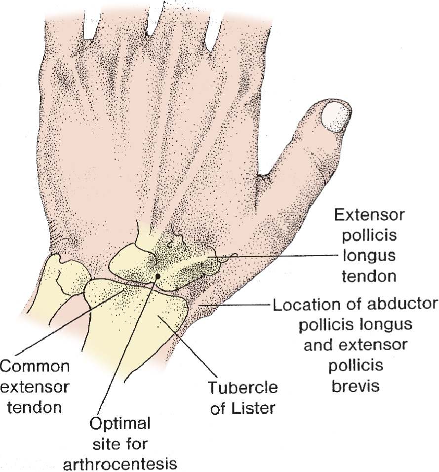
![]() Palpate the dorsal radial tubercle (Lister tubercle) and the extensor pollicis longus tendon as it courses over the distal radius
Palpate the dorsal radial tubercle (Lister tubercle) and the extensor pollicis longus tendon as it courses over the distal radius
![]() Palpate the depression that is distal to the tubercle and on the ulnar side of the extensor carpi radialis brevis tendon
Palpate the depression that is distal to the tubercle and on the ulnar side of the extensor carpi radialis brevis tendon
![]() Ulnocarpal Approach
Ulnocarpal Approach
![]() Flex the wrist 20 degrees and palpate the depression between the ulnar styloid process and pisiform bone
Flex the wrist 20 degrees and palpate the depression between the ulnar styloid process and pisiform bone
![]() Approach may be problematic due to multiple tendons travel through this region
Approach may be problematic due to multiple tendons travel through this region
TECHNIQUE
![]() Patient Preparation
Patient Preparation
![]() Confirm landmarks—mark the needle insertion point if needed
Confirm landmarks—mark the needle insertion point if needed
![]() Sterilize the area where the needle will be inserted with povidone–iodine solution or comparable skin antiseptic
Sterilize the area where the needle will be inserted with povidone–iodine solution or comparable skin antiseptic
![]() Wipe injection site with alcohol to avoid introduction of iodine solution into the synovium
Wipe injection site with alcohol to avoid introduction of iodine solution into the synovium
![]() Drape the area with sterile towels
Drape the area with sterile towels
![]() Place the wrist in neutral, relaxed position
Place the wrist in neutral, relaxed position
![]() Apply gentle traction and ulnar deviation to the hand to open the joint space
Apply gentle traction and ulnar deviation to the hand to open the joint space
![]() Analgesia
Analgesia
![]() Use a 25-gauge needle to infiltrate injection site with lidocaine with epinephrine
Use a 25-gauge needle to infiltrate injection site with lidocaine with epinephrine
![]() Anesthetize the subcutaneous tissue and a track toward the joint
Anesthetize the subcutaneous tissue and a track toward the joint
![]() Avoid entering the joint space if synovial fluid analysis is desired
Avoid entering the joint space if synovial fluid analysis is desired
![]() Aspiration
Aspiration
![]() Use a 22-gauge needle attached to a 5- or 10-mL syringe
Use a 22-gauge needle attached to a 5- or 10-mL syringe
![]() For the radiocarpal approach, direct the needle just distal to the border of the distal radius
For the radiocarpal approach, direct the needle just distal to the border of the distal radius
![]() Insert the needle in the depression on the ulnar side of the extensor carpi radialis brevis tendon and between the distal radius and lunate bone (FIGURE 60.1)
Insert the needle in the depression on the ulnar side of the extensor carpi radialis brevis tendon and between the distal radius and lunate bone (FIGURE 60.1)
![]() For the ulnocarpal approach direct the needle between the distal border of the ulnar styloid process and the pisiformis bone (FIGURE 60.2)
For the ulnocarpal approach direct the needle between the distal border of the ulnar styloid process and the pisiformis bone (FIGURE 60.2)
![]() Provide negative pressure on the syringe plunger as the needle is inserted in the joint cavity
Provide negative pressure on the syringe plunger as the needle is inserted in the joint cavity
![]() Easy aspiration of fluid confirms proper needle position
Easy aspiration of fluid confirms proper needle position
![]() Withdraw needle, apply pressure, then apply clean dressing
Withdraw needle, apply pressure, then apply clean dressing
COMPLICATIONS
![]() Iatrogenic infection
Iatrogenic infection
![]() Increased pain
Increased pain
![]() Localized bleeding
Localized bleeding
![]() Reaccumulation of effusion
Reaccumulation of effusion
![]() Injury to articular cartilage if proper technique is not utilized
Injury to articular cartilage if proper technique is not utilized

FIGURE 60.1 Radiocarpal arthrocentesis. (From Simon RR, Brenner BE. Emergency Procedures and Techniques. 4th ed. Philadelphia, PA: Lippincott Williams & Wilkins; 2002:239, with permission.)
Stay updated, free articles. Join our Telegram channel

Full access? Get Clinical Tree


