 Used to provide anesthesia in distribution of median, ulnar, and/or radial nerves for the treatment of complex soft-tissue or bony injuries of the hand
 Irrigation of deep abrasions with embedded debris
 Extensive or complex laceration repair
 Burn injury pain control
 Incision and drainage of abscess
 Fracture/dislocation reduction
 Traumatic amputation
CONTRAINDICATIONS
 Overlying cellulitis at site of anticipated injection
 Relative Contraindications
 Simple laceration or injury that can be easily and adequately anesthetized with local infiltration or digital block
COMPLICATIONS
 Hematoma formation and/or vascular injury
 Nerve injury
 Infection
 Allergic reaction
 General Basic Steps
 Palpate landmarks
 Sterile prep of skin
 Inject anesthetic
 Consider sensory branches
ULNAR NERVE BLOCK
 Landmarks
 Flexor carpi ulnaris tendon, pisiform bone, ulnar artery, proximal wrist crease (FIGURE 82.1)
 Ulnar nerve splits into dorsal and palmar branches approximately 5 cm proximal to the wrist crease
 Palmar branch runs between the flexor carpi ulnaris tendon and the ulnar artery at the level of the proximal wrist crease
 Technique
 Patient Preparation
 Place the hand comfortably on bedside procedure table with palmar surface up
 Prepare wrist site in standard sterile manner (povidone–iodine solution or chlorhexidine)
 Have patient flex wrist against resistance to accentuate landmarks
 Identify and mark the flexor carpi ulnaris tendon from its insertion site at the pisiform bone to the ulnar nerve branch point (approximately 5 cm proximal to wrist crease)
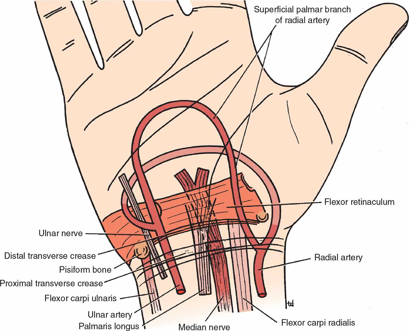
FIGURE 82.1 Surface anatomy of the wrist region. (From Snell RS. Clinical Anatomy. 7th ed. Philadelphia, PA: Lippincott Williams & Wilkins; 2004:534, with permission.)
 Injection
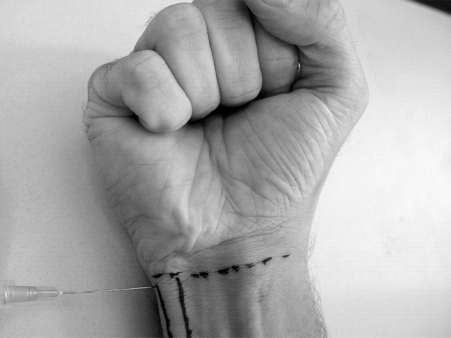
 Approach the wrist medially and insert a small-bore (25-gauge) needle beneath the flexor carpi ulnaris tendon at the level of the proximal wrist crease (FIGURE 82.2)
 Inject approximately 2 mL of anesthetic solution (lidocaine/bupivacaine) beneath the tendon at its radial border
 Aspirate before injecting anesthetic to ensure that ulnar artery has not been inadvertently entered
 To anesthetize the dorsal branch of the ulnar nerve use the same initial insertion site and technique. However, redirect needle 3 to 5 cm proximally toward the branch point.
 Inject approximately 5 mL of anesthetic solution beneath the tendon at this branch point
Related posts:



Stay updated, free articles. Join our Telegram channel

Full access? Get Clinical Tree


