Tissue trauma accounts for significant morbidity and financial concern. Treatment of chronic wounds conservatively costs the U.S. health care system $50 billion annually.1 The incidence and prevalence of acute and chronic wounds vary with populations, geographic and demographic status, and general medical conditions. Acute wounds consist of lacerations, abrasions, avulsions, crush injuries, puncture wounds, insect or mammalian bites, traumatic or surgical wounds, burns, and skin tears. Chronic wounds consist of pressure ulcers, venous and arterial ulcers, diabetic foot ulcers, and nonhealing surgical or traumatic wounds.
Pressure and Venous Ulcers
An estimated 2.5 million pressure ulcers are treated each year in U.S. acute care facilities alone, and the cost of treating them ranges from $11 billion to $17.2 billion per year.2 Development of a pressure ulcer increases the length of stay for hospitalized patients, and increases the mortality rate significantly.2
Venous ulcers account for 80% to 90% of ulcers found on the lower leg.3 Approximately 26% to 28% of healed ulcers will recur within 1 year.3 The annual cost of treating venous stasis ulcers to the U.S. health care system is $1.9 billion to $3.5 billion.3 Arterial ulcers occur in a large percentage of patients with peripheral arterial disease. These ulcers are particularly difficult to heal as a result of poor perfusion, and revascularization is often needed to promote reperfusion and the hope of healing the ulcer.
Diabetic Foot Ulcers
There are more than 20 million people in the United States with diabetes, of whom 10% to 15% are at risk for ulceration.2 Of all nontraumatic lower limb amputations in the United States, 50% to 75% are diabetes related.2 Amputation and foot ulceration are the most common consequences of diabetic neuropathy and major causes of morbidity and disability in people with diabetes. People with diabetes are at higher risk for development of peripheral artery disease, which significantly increases the risk of amputation. The risk of ulcers or amputations also increases in people who have had diabetes for more than 10 years, are male, or have poor glucose control; risk is also increased with cardiovascular, retinal, or renal complications (Boxes 69-1 and 69-2).4
Box 69-1
Foot Care Recommendations for Patients with Diabetes
From American Diabetes Association: Standards of medical care in diabetes—2010, Diabetes Care 33(Suppl 1):S11-S61, 2010.
Surgical Wounds
Surgical wounds are often treated in the outpatient setting secondary to the rising costs of acute care management and reimbursement concerns. Wound dehiscence or infection can affect healing by secondary intention and may require tertiary healing, with the health care provider and home health nurse managing the care of these difficult-to-heal wounds.5,6 A broad understanding of wound healing physiology and general management principles will assist the provider in facilitating maximum wound repair.
Classification of Wounds
Classification of wounds is unique to wound type, and reference to established classification systems is recommended.
Wounds involving only the epidermal layer are classified as superficial or partial thickness. Examples include simple lacerations, skin tears, first-degree burns, abrasions, and shallow ulcerations. These wounds usually heal quickly and require the least intervention. Full-thickness wounds involve the epidermis and dermis and may extend through subcutaneous tissue into muscle and bone. Examples include deep lacerations, second- and third-degree burns, various types of ulcers, and surgical or traumatic wounds.
A pressure ulcer is a localized injury to the skin or underlying tissue and is staged in accordance with the National Pressure Ulcer Advisory Panel’s staging definitions. Pressure ulcer staging includes deep tissue injury, stages I to IV, and unstageable (Box 69-3). Burns are classified as first, second, or third degree (Box 69-4) and require a unique approach that often requires referral to a burn specialist (Box 69-5).
Box 69-3
Pressure Ulcer Staging (NPUAP/EPUAP Guidelines)
Definition
A pressure ulcer is localized injury to the skin and/or underlying tissue, usually over a bony prominence, as a result of pressure, or pressure in combination with shear and/or friction. A number of contributing or confounding factors are also associated with pressure ulcers; the significance of these factors is yet to be elucidated.
Purple or maroon localized area of discolored intact skin or blood-filled blister caused by damage of underlying soft tissue from pressure and/or shear. The area may be preceded by tissue that is painful, firm, mushy, boggy, warmer or cooler as compared with adjacent tissue.
Further description: Deep tissue injury may be difficult to detect in individuals with dark skin tones. Evolution may include a thin blister over a dark wound bed. The wound may further evolve and become covered by thin eschar. Evolution may be rapid, exposing additional layers of tissue even with optimal treatment.
Stage I
Intact skin with nonblanchable redness of a localized area, usually over a bony prominence. Darkly pigmented skin may not have visible blanching; its color may differ from the surrounding area.
Further description: The area may be painful, firm, soft, warmer or cooler as compared with adjacent tissue. Stage I wounds may be difficult to detect in individuals with dark skin tones. May indicate “at-risk” persons (a heralding sign of risk).
Stage II
Partial-thickness loss of dermis manifesting as a shallow open ulcer with a red or pink wound bed, without slough. May also manifest as an intact or open or ruptured serum-filled blister.
Further description: Shiny or dry shallow ulcer without slough or bruising.* This stage should not be used to describe skin tears, tape burns, perineal dermatitis, maceration, or excoriation.
Stage III
Full-thickness tissue loss. Subcutaneous fat may be visible, but bone, tendon, or muscle is not exposed. Slough may be present but does not obscure the depth of tissue loss. May include undermining and tunneling.
Further description: The depth of a stage III pressure ulcer varies by anatomic location. The bridge of the nose, ear, occiput, and malleolus do not have subcutaneous tissue, and stage III ulcers can be shallow. In contrast, areas of significant adiposity can develop extremely deep stage III pressure ulcers. Bone or tendon is not visible or directly palpable.
Stage IV
Full-thickness tissue loss with exposed bone, tendon, or muscle. Slough or eschar may be present on some parts of the wound bed. Often includes undermining and tunneling.
Further description: The depth of a stage IV pressure ulcer varies by anatomic location. The bridge of the nose, ear, occiput, and malleolus do not have subcutaneous tissue, and these ulcers can be shallow. Stage IV ulcers can extend into muscle and/or supporting structures (e.g., fascia, tendon, or joint capsule), making osteomyelitis possible. Exposed bone or tendon is visible or directly palpable.
Unstageable
Full-thickness tissue loss in which the base of the ulcer is covered by slough (yellow, tan, gray, green, or brown) and/or eschar (tan, brown, or black) in the wound bed.
Further description: Until enough slough and/or eschar is removed to expose the base of the wound, the true depth, and therefore stage, cannot be determined. Stable (dry, adherent, intact without erythema or fluctuance) eschar on the heels serves as “the body’s natural (biologic) cover” and should not be removed.
From National Pressure Ulcer Advisory Panel (NPUAP) and European Pressure Ulcer Advisory Panel (EPUAP): Pressure ulcer prevention and treatment: clinical practice guideline, Washington, DC, 2009, National Pressure Ulcer Advisory Panel.
Box 69-4
Burn Classification
First-Degree (Superficial or Epidermal) Burns
These burns involve only the epidermis. They do not blister, but are red and quite painful. Over 2 to 3 days the erythema and the pain subside. By about day 4, the injured epithelium peels away from the newly healed epidermis underneath, a process that is commonly seen after sunburn.
Second-Degree (Partial-Thickness) Burns
Partial-thickness burns involve the epidermis and portions of the dermis and can be clinically categorized as either superficial partial-thickness or deep partial-thickness burns. Superficial partial-thickness burns characteristically form blisters between the epidermis and dermis. Because blistering may not occur for some hours after injury, burns that initially appear to be only epidermal in depth (first degree) may be determined to be partial-thickness burns 12 to 24 hours later. Most superficial partial-thickness burns heal spontaneously in less than 3 weeks, and do so typically without functional impairment or hypertrophic scarring. Second-degree burns often accumulate a layer of fibrinous exudate and necrotic debris on the surface, which may predispose the wound to heavy bacterial colonization and delayed wound healing, in addition to making more difficult the determination of wound depth by visual inspection.
Deep partial-thickness burns extend into the lower layers of the dermis. They possess characteristics that are distinctly different from superficial or mid-dermal partial-thickness burns. If infection is prevented and spontaneous healing is allowed to progress, these burns will heal in 3 to 9 weeks. However, they invariably cause considerable scar formation. Even with active physical therapy throughout the healing process, hypertrophic scarring is common and joint function is usually impaired. These burns are best treated by excision and grafting. For the patient, a partial-thickness burn that fails to heal within 3 weeks is functionally and cosmetically equivalent to a full-thickness injury.
Third-Degree (Full-Thickness) Burns
Full-thickness burns involve all layers of the dermis and often injure underlying subcutaneous adipose tissue as well. Burn eschar is structurally intact but dead and denatured dermis. Over days and weeks, if left in situ, eschar separates from the underlying viable tissue, leaving an open, unhealed bed of granulation tissue. Without surgery, these burns can heal only by wound contracture with epithelialization from the wound margins. Some full-thickness burns involve not only all layers of the skin, but also deeper structures such as muscle, tendon, ligament, and bone, and are classified as deep full-thickness or fourth-degree burns. Grafting may use autologous skin grafts or biologic dressings and skin substitutes or both. (Excision and grafting using biologic dressings or skin substitutes permits closure of extensive burns in stages, with autografting done at a later date; see detailed discussion elsewhere in this chapter.) Deep full-thickness burns may require amputation or closure with alternative techniques (such as adjacent tissue transfer or microvascular procedures).
From Kagan RJ, Peck MD, Ahrenholz DH, et al: Surgical management of the burn wound and use of skin substitutes: an expert panel white paper. J Burn Care Res 34(2):e60-e79, 2013.
Box 69-5
Burn Center Referral Criteria
A burn center may treat adults, children, or both. Burn injuries that should be referred to a burn center include the following:
From American College of Surgeons, Committee on Trauma: Guidelines for the operation of burn centers. In Resources for optimal care of the injured patient, Chicago, Ill, 2006, American College of Surgeons.
Skin tears are common in frail older adults, often occurring during routine daily activities such as washing and dressing. The shearing and friction forces against frail skin cause the tear, separating the epidermis from the dermis (partial thickness) or the dermis from underlying structures (full thickness).
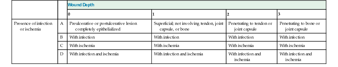
Arterial and venous ulcers are classified as partial or full thickness; the diabetic foot ulcer is typically graded by one of two classification systems, the Wagner and the University of Texas classifications. The Wagner system uses grades 0 to 5 to assess wound depth (Table 69-1). The University of Texas system uses a matrix of grades and scales to assess the wound’s depth and presence of infection or ischemia (Table 69-2).7,8
Wound healing begins at the time of injury and often proceeds over a period of several months through the stages of inflammation, proliferation, and remodeling. Inflammation, which begins at the time of injury, is an essential first step in wound healing to provide local vasospasm and initiation of the clotting process. Neutrophils, oxygen, and nutrients are transported to the wound site, and proliferation begins. In this phase, epithelial cells migrate over the surface of the wound, collagen synthesis begins, and the wound begins to contract. Remodeling occurs during the next several months, with organized layering of type I collagen providing improved tensile strength.2,7,9
Wound healing is affected by many internal and external factors. Internal factors include age, preexisting comorbidities (e.g., diabetes mellitus, cardiovascular disease, autoimmune disorders), perfusion, oxygenation, nutrition, hydration, and some medications (especially steroids, immunosuppressants, and chemotherapeutic drugs). External factors include pressure, friction, shear, contamination (with bacteria, debris, or necrotic tissue), and wound environment (pH, moisture).2,7
Stages of wound healing may be interrupted by changes in the internal and external wound healing factors. Two common examples are the occurrence of anemia during wound healing, which slows the healing response as a result of decreased oxygenation, and pressure exerted over the site, which decreases perfusion and prolongs or delays wound healing.7
Surgical wounds heal by primary, secondary, or tertiary intention. Primary intention implies that the wound edges are approximated and sutured, stapled, taped, or glued. Secondary intention implies that the wound edges are not approximated, usually because of failed primary intention (dehiscence) or infection. Secondary intention healing is prolonged and results in significant scarring. Delayed primary intention, or tertiary intention, refers to wounds that were not initially closed (usually because of infection, contamination, or wound stress) and are closed after some secondary intention healing has occurred.
Only gold members can continue reading. Log In or Register to continue