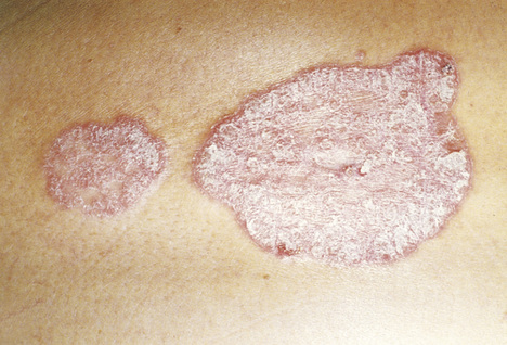
Richard M. Prior Psoriasis is an inflammatory papulosquamous eruption characterized by well-circumscribed erythematous macular and papular lesions with loosely adherent silvery white scale. It is a chronic, unpredictable disorder that is characterized by remissions and exacerbations throughout the life span. From 1% to 3% of the population is affected by psoriasis, or around 7.4 million Americans, with 25% to 45% of cases beginning after the age of 10 years. First episodes often appear in young adulthood, but they can appear later in life as well. Stress, anxiety, and illness often precede flares. Time lost from school and work as well as the emotional and financial constraints on families mandate effective and convenient treatments. Symptoms can be treated; however, as yet there is no cure. Remissions are common and can last for short periods or years, with progression to arthritis in about 30% of cases. A genetic component appears to exist in this disorder; thus a familial tendency can increase risk.1,2 Patients with psoriasis score poorly on quality-of-life (QOL) measures. Patients are troubled by the appearance of the lesions and pruritus associated with the disease. Those with psoriatic arthritis often have disability and pain. Over 80% of those with the disorder report that psoriasis often affects their emotional state and decreases their satisfaction with life. Psoriasis and psoriatic arthritis can have negative economic effects on those with the disorder; more than 92% of unemployed patients attribute their lack of a job solely to their disease.3 Psoriasis should be viewed as a systemic disorder that causes additional morbidity in those affected. Patients with psoriasis are more prone to inflammatory bowel disease and cardiovascular disease. They are likely to be overweight, hypertensive, diabetic, and dyslipidemic when psoriasis symptoms are present. In addition, these patients are more likely to experience alcohol dependence and depression.4 Psoriasis is a chronic, inflammatory, autoimmune disorder characterized by dermal hyperproliferation that develops in response to T-cell infiltration into the skin and overexpression of multiple cytokines, including interferon, tumor necrosis factor (TNF), and interleukin-23 (IL-23). T cells are activated and produce an inflammatory response that results in the hyperproliferation of keratinocytes. Psoriasis lesions often contain 30 times the number of keratinocytes as normal skin.1 Psoriasis is known to have strong genetic associations. Those who have first-degree family members with the disorder are often affected as well. There is an increased incidence of psoriasis in monozygotic twins.1 In psoriasis, scaly papules and plaques form and collect on skin surfaces in well-demarcated lesions (Fig. 62-1). The lesions have an erythematous base with silvery white plaques that are adherent. The dermis is highly vascular, and tiny bleeding points are revealed if the scales are removed (Auspitz sign). Common sites for these lesions include the elbows, knees, scalp, genitals, and intergluteal cleft. In contrast to adult psoriasis, childhood psoriasis often involves the face. Many patients exhibit concomitant nail dystrophies, including pitting, yellowing of the distal portion (oil drop sign), separation of the nail plate (onycholysis), and thickening of the entire nail (hyperkeratosis).5 Cutaneous trauma can induce psoriasis 1 to 3 weeks after injury. This isomorphic response, also known as the Koebner phenomenon, occurs in a linear fashion along the lines of a scratch, abrasion, sunburn, or pressure. Discrete, scaly, “raindrop” plaques that are smaller than 1 cm, begin on the trunk, and spread to the extremities, sparing the palms and soles, are indicative of guttate psoriasis. Guttate psoriasis is occasionally seen after a streptococcal infection and is most common in adolescents. These patients are likely to develop psoriasis vulgaris (common, plaquelike psoriasis) later in life.5 Erythroderma and pustular psoriasis are more serious forms of the disease. They are most common in patients older than 50 years and may be precipitated by infection, withdrawal of systemic steroids, emotional stress, or severe illness. Erythrodermic forms generally appear over a large portion of the body and can be precipitated by various treatments themselves.5 Although most psoriatic lesions are asymptomatic, itching is variable. However, picking and scratching of the lesions can produce the Koebner response, and the lesions worsen. Skinfold lesions tend to itch more than common plaquelike lesions. The axilla, intramammary folds, groin, buttocks, and genitals are common sites for intense itching, or inverse psoriasis. The bright red appearance of the lesions and affinity for dark, moist folds can make distinguishing inverse psoriasis from Candida infections difficult based on appearance alone. Psoriatic arthritis is a seronegative spondyloarthropathy that affects approximately 10% to 15% of the population with psoriasis. It is characterized by monoarthritis, often causing joint effusions; pain at the insertion point of tendons to bone (enthesitis); swelling of the fingers and toes (dactylitis); and changes to the nails (including pitting and splitting). In a small percentage of patients, psoriatic arthritis precedes the appearance of skin symptoms. The presence of silvery scales on red, erythematous plaques is characteristic; therefore the diagnosis is usually based on presentation. However, biopsy is useful in pustular cases, and nail cultures differentiate fungal disease. Uric acid levels may be elevated in psoriasis as well as in gout. Psoriatic arthritis is diagnosed clinically based on symptom scoring models, as there is no diagnostic laboratory test. As many as half of those with psoriatic arthritis may be HLA-B27 positive. Almost all patients with psoriatic arthritis are rheumatoid factor negative. Many patients who develop erosive arthritis have radiographic findings in advanced stages of the disease. In children, the plaques of psoriasis are thinner and less scaly than in adults with psoriasis and are often confused with seborrhea, atopic dermatitis, and diaper dermatitis. Seborrhea on the scalp tends to be patchy, red, and a bit oilier in appearance. Psoriasis is more plaquelike, with thick scales. Psoriasis typically appears on extensor surfaces, whereas atopic dermatitis is found on most flexor surfaces. Lichen planus papules have more of a purple hue, and patients exhibit Wickham striae (lacy, reticular, crisscrossed whitish lines) on many lesions. Flat warts do not have scale on the surface. Guttate psoriasis is often confused with pityriasis rosea; however, it lacks the characteristic herald patch, and the scale is thicker and more diffuse in psoriasis. Changes in the nails are often confused with onychomycosis. Culture for the presence of fungus will help establish the diagnosis. Yellow discoloration is common in both fungal and psoriatic changes, as is nail separation. The nails in psoriasis are not well formed because debris collects underneath, again because of rapid shedding of the skin layers. This debris leads to failure in the integrity of the nail and onycholysis. Additional diagnoses to be considered are gout, pseudogout, reactive arthritis, syphilis, squamous cell carcinoma, nummular eczema, and lichen simplex chronicus.
Psoriasis
Definition and Epidemiology
Pathophysiology
Clinical Presentation and Physical Examination
Diagnostics
Differential Diagnosis
Psoriasis
Chapter 62