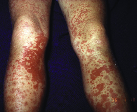
Joanne Sandberg-Cook Purpura is a hemorrhaging into the skin. The size of the bleeding vessel determines the size of the lesion, which in turn may provide clues to the cause. Petechiae are lesions less than 3 mm (1/10 inch) in diameter; these indicate capillary bleeding. Lesions ranging from 3 mm to 1 cm (1/10 to 4/10 inch) are often referred to as purpura. Lesions larger than 1 cm are referred to as ecchymoses. All show a predilection for the limbs. Purpura is divided into two groups: inflammatory (palpable) and noninflammatory. Noninflammatory purpura is further divided into hemostatic defects, nonpalpable purpura, and nonhemostatic defects (vascular purpura).1 Purpura is characterized by an extravasation of red blood cells into the dermis from small cutaneous vessels. Hemosiderin or hematoidin may be present if the purpura is chronic; this causes a characteristic red or brown discoloration. Purpura may be oval or round or irregularly outlined; it may be flat or raised (palpable) as a result of edema or induration.1 Palpable purpura consists of raised, erythematous lesions that do not blanch when the skin is pressed with a glass slide. Dilated superficial capillaries, in which the blood remains confined within the vessels, do blanch when pressed, thereby distinguishing them from true purpura. Extravasation of blood from the vessel depends on the integrity of the blood vessel, which in turn depends on the strength of the vessel, the transmural pressure gradient that drives blood out of the vessel, and the competence of the mechanism that combats the basal level of vascular trauma.1 Because purpura is a symptom of many systemic diseases, these lesions seldom are seen without other symptoms. A review of systems should include an inquiry into other bleeding sites, abnormally heavy menstrual bleeding, trauma, recent infection (including sexually transmitted diseases), exposure to ticks or a tick bite, and recent travel to areas where Rocky Mountain spotted fever, dengue fever, or Lyme disease is endemic or epidemic. A complete medication history (including over-the-counter medications and allergies) should be taken. Any history of autoimmune disease or other serious illnesses, such as leukemia or lymphoma, should be noted. Recent complaints of fever, chills, arthralgias, and myalgias should be noted. The skin is the focus of the physical examination. The size, location, and shape of the lesions should be documented. Bullae and ulcerations can develop within any lesion larger than petechiae.2 Lesions should be palpated for swelling (palpable purpura) or flatness against the skin. Palpable purpura is usually associated with inflammation of the vessel (Fig. 63-1; see Chapter 220). A glass slide pressed against the lesion determines whether it is blanchable, thereby differentiating it from erythema or dilated superficial capillaries.1 Excoriation may imply pruritus. The remainder of the general examination includes an oral examination to look for lesions of the gums or tongue and a joint examination to look for swelling, inflammation, or deformities that would suggest connective tissue disease. Fever, nuchal rigidity, organomegaly, or a new heart murmur may imply serious systemic disease or infection. Observations of weight, nutritional status, or skin turgor may suggest nutritional deficiencies. Evidence of trauma (healing bruises, fractures) may indicate ongoing trauma as a cause. Laboratory studies help differentiate between inflammatory and noninflammatory purpura. (Inflammatory purpura [vasculitis] is discussed in Chapter 220.) A complete blood count (CBC) with a platelet count (not an estimate) is most helpful. An erythrocyte sedimentation rate (ESR) or C-reactive protein (CRP) level can be beneficial in excluding an inflammatory cause. Bleeding time, platelet count, prothrombin time (PT), partial thromboplastin time (PTT), and international normalized ratio (INR) will determine the presence of coagulopathies. Blood urea nitrogen (BUN), creatinine, and liver function tests (LFTs) are necessary to exclude organ disease. Immune studies to exclude autoimmune diseases such as lupus, rheumatoid arthritis, cryoglobulinemias, or scleroderma may be indicated, depending on other physical findings and symptoms. Infectious disease screening would include screening for hepatitis B and C, human immunodeficiency virus (HIV) infection, and tuberculosis.2
Purpura
Definition and Epidemiology
Pathophysiology
Clinical Presentation
Physical Examination
Diagnostics
Purpura
Chapter 63