Vascular Access Issues and Procedures

Chapter 11


Vascular Access Issues and Procedures image


Obtaining access to the vasculature is an essential skill for clinicians in intensive care units (ICUs). Arterial catheterization is used to continuously monitor blood pressure and to obtain blood for arterial blood gas analysis and other laboratory tests. Central venous catheterization permits measurement of central venous pressures (CVPs); rapid infusion of fluids; and central administration of certain agents, such as calcium, potassium, vasoactive agents, or hyperalimentation solutions. This chapter begins with a brief overview of the use of ultrasound to guide vascular access, followed by a description of the indications, techniques, and risks of cannulation of arteries, veins, and pulmonary artery.


Bedside Ultrasonography in the ICU


Technical Considerations


Before beginning any procedure with ultrasound guidance, the operator must have familiarity with the equipment. This includes, but is not limited to, knowledge of the various types of probes, the specific ultrasound machine in use, and appropriate knowledge of the anatomy to be interrogated. Additionally, an understanding of ultrasound physics and image interpretation (as well as knowledge of ultrasound artifacts) is necessary. Vascular access typically employs a linear array transducer. This probe can be recognized by the flat front on the transducer. It will produce a square image on the screen, in contrast to the wedge-shaped image of a phased-array probe. On one side of the probe is an indicator that corresponds to the marker on the left-hand side of the display screen.


The depth of the display and image gain should be set so that the structure of interest is in the middle of the screen and tissue density is appropriately shaded, respectively.


Methodology


Ultrasound can be used in both static and dynamic capacities to aid in vascular access. Static ultrasound is the use of imaging to localize, characterize, and mark the vessel prior to beginning the procedure. In contrast, dynamic ultrasound uses imaging to visualize the needle and the vessel in real time during the procedure and to guide puncture of the vessel. It is optimal to apply both techniques: (1) assess vessel location and patency prior to preparing the patient and (2) perform image the procedure using dynamic ultrasound guidance.


Vessels can be imaged in a transverse or longitudinal fashion. Transverse imaging allows for easy identification of surrounding structures (e.g., carotid artery during internal jugular cannulation). This technique, however, requires greater skill than longitudinal imaging as it is often difficult to differentiate between the tip and shaft of the needle. Without knowing where the tip of the needle is at all times, ultrasound becomes useless and can be dangerous as it may give a false sense of security. Longitudinal imaging allows one to visualize the needle throughout its length and as it pierces the vessel. This, however, requires greater coordination as the operator must ensure that the ultrasound plane of imaging does not move during needle insertion.


After adequate images are obtained, the skin is entered with the needle above the vessel. It often takes one hand to hold the skin taught and the other to pierce the skin with the needle. Next, using the dominant hand to guide the needle and the nondominant hand to hold the probe, the needle can be visualized and advanced. A needle guide to align the probe with needle can be helpful for beginners but is not necessary. The needle appears as a hyperechoic (white) structure. If transverse imaging is utilized, it is helpful to gently fan or rock the probe to bring the tip of the needle into and out of the plane of the image. This allows one to determine if he or she is seeing the shaft or the tip of the needle on the screen.


Arterial Catheterization


Indications


Measuring blood pressure with an intra-arterial catheter is preferable to non-invasive methods when there is a need for frequent blood pressure readings. Each of the determinants of blood pressure (blood volume, systemic vascular resistance, and cardiac contractility) can change rapidly under certain circumstances. For example, effective blood volume changes minute by minute in response to major gastrointestinal bleeding or to large increases in intrathoracic pressure. Systemic vascular resistance can also vary rapidly with changes in patient temperature (e.g., rewarming after therapeutic hypothermia) or in response to administration of vasoactive drugs. Drugs that depress cardiac contractility or rate can also cause rapid decreases in blood pressure. Newer forms of hemodynamic monitoring that employ analysis of the arterial waveform or variations in arterial pulse pressure to determine cardiac output or predict fluid responsiveness also require arterial catheterization. In addition, arterial access may be required to sample arterial blood to determine pH, Pao2, and Paco2 frequently in patients with acute respiratory failure or acid-base disorders. Finally, the need for frequent phlebotomy may warrant insertion of an arterial catheter for patient comfort.


Sites


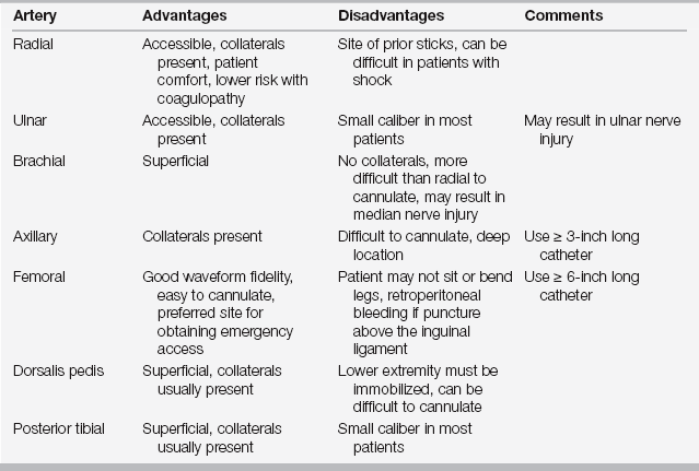
A number of superficial arteries are used for arterial catheterization in the adult, including the radial, ulnar, brachial, axillary, femoral, dorsalis pedis, and posterior tibial arteries (Table 11.1). Although the radial artery is the most commonly used site, alternative sites may be useful in certain situations. For example, when the radial artery is not palpable, the femoral artery is an acceptable alternative. Although some ICU clinicians perform an Allen test before radial artery cannulation to document the presence of collateral circulation (via the ulnar artery), many others do not. The medical literature, including large series of patients receiving radial artery cannulation, is equivocal regarding any benefit to including this test prior to cannulation. In addition, there is no consensus on the definition or significance of an abnormal test. Clearly, after arterial catheterization of any site, the distal extremity must be monitored for signs and symptoms of ischemia. If there is concern for ischemia, the arterial catheter should be removed and a vascular surgeon should be consulted for immediate evaluation.


As with all procedures on ICU patients, the operator should observe standard safety measures prior to starting the procedures, including a “time out” or “pause” for safety, and document the pause (see Chapter 109). At a minimum, this should confirm that the following are correct: (1) the patient, (2) the procedure, and (3) the extremity and laterality (right versus left side). To perform vascular access using dynamic ultrasound, the ultrasound should be positioned in a manner that allows the practitioner easy viewing access without strain or discomfort. After the site is prepped in a standard and sterile manner, the operator dons hat, gown, and gloves. The sterile area should be draped, and ultrasound gel is placed inside a sterile plastic sleeve. An assistant then lowers the ultrasound probe into the sleeve taking care to avoid contamination of the operator, the sterile field, or the outer surface of the sleeve. The sleeve should be extended so that it covers both probe and cable, allowing the operator mobility without concern for impairing the sterility of the field. Sterile gel is then placed on the patient over the site of interest.


The probe can now be used to define the internal anatomy. Most fluid, including blood, appears black and hypoechoic. Veins are readily identified, as they are typically larger and more compressible than arteries. Additionally, if color flow Doppler is utilized, pulsatile flow is usually not seen in veins. Note that reliance solely on this latter feature may cause confusion in patients with severe tricuspid insufficiency. Additionally, in patients who are severely hypotensive, arteries may exhibit easy compressibility, making it more difficult to differentiate them from veins.


Common upper extremity alternatives to the radial artery include the brachial and axillary arteries. The brachial artery is superficial, readily palpated, and easily imaged by ultrasound; however, it has the disadvantage of being an anatomic end-artery that lacks a collateral circulation. Consequently, some intensivists prefer the axillary artery because it has a collateral circulation; however, the axillary artery can be more difficult to cannulate as well as to repair surgically in case of dissection or thrombosis.


In the lower extremity, the femoral artery is the preferred site for arterial cannulation. It is easy to access, and its arterial waveform usually demonstrates good fidelity. Patients with indwelling femoral catheters should not be mobilized from bed to chair to prevent injury to the artery. In addition, there are serious complications that can result from the procedure itself, including retroperitoneal bleeding (when the artery is punctured above the inguinal ligament) and atheromatous emboli in patients with diffuse atherosclerosis. The dorsalis pedis artery is an alternative in the lower extremity because it generally has good collateral flow.


Insertion Methods


There are two common approaches to cannulating an artery in the ICU: direct cannulation of the vessel and the modified Seldinger technique. The direct approach involves insertion of the needle into the vessel lumen until arterial blood is identified. At this point the catheter is gently threaded over the needle into the artery. The direct approach can be used for catheterization of smaller vessels; however, it is often more difficult to perform and is not suggested for inexperienced operators.


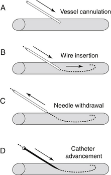
The modified Seldinger technique is a multistage procedure (Figure 11.1). The first step involves insertion of the needle into the vessel lumen. Ultrasound guidance may be helpful during this step. The second step involves passing a thin guide wire through the needle into the lumen. After the wire is in the vessel lumen, the introducer needle is removed while the operator maintains control of the guide wire. Next, a plastic catheter is threaded over the wire into the vessel. The wire is now removed from the catheter lumen. Success is confirmed by the return of pulsatile blood from the catheter. Care should be taken to avoid traumatizing the vessel. The wire typically has a flexible, often J-shaped, tip and only this end of the wire should enter the vessel. The wire should never be advanced if resistance is met. Finally, a complication of this technique is inadvertent loss of the wire into the vessel, which can be avoided by maintaining control of some portion of the wire throughout the procedure.


The traditional modified Seldinger technique is used for larger vessels such as the femoral artery; however, two variations are helpful, particularly for smaller vessels such as the radial artery. The first is use of arterial line kits that have an integrated needle, catheter, and guide wire. The kits are used in a similar fashion as described previously, except once the vessel is entered with needle, the wire is advanced, and the catheter is simply advanced over the needle and into the vessel.


The second method utilizes a separate guide wire. After puncturing the skin with the catheter and visualizing arterial blood in the reservoir, the catheter and needle are advanced slightly further so that the back wall of the artery is punctured (the so-called through-and-through technique). The needle is then removed. The catheter is very slowly withdrawn into the vessel. When pulsatile blood flow is again obtained, a small guide wire is advanced through the catheter into the vessel lumen. Finally, the catheter is advanced over the wire into the vessel.


Complications


Risks of arterial catheterization include infection, thrombosis, and hemorrhage. The risk of arterial catheter-related bloodstream infection is controversial, but it is certainly lower than the risk of central venous catheter infection. There are minimal data to support the practice of routine changes in cannula and/or cannulation site and such a practice is discouraged. While bloodstream infection from arterial catheters is uncommon, colonization of the hubs of their three-way stopcocks by bacteria, such as coagulase-negative staphylococcal species, is common. Consequently, one should never use an arterial catheter to obtain blood cultures except at the time of its first insertion.


Thrombosis of arterial lines is most likely to occur when the cannulated vessel is anatomically abnormal (calcified, narrowed) or if there is circulatory shock and impaired blood flow. Central (retrograde) embolization of air bubbles or thrombi with serious consequences can occur if an image arterial catheter is flushed vigorously.


Peripheral Venous Catheterization


Peripheral intravenous (IV) catheters are present in all patients in the ICU. Insertion is typically performed using a catheter-over-needle technique. Ultrasound has been shown to be helpful when venous cannulation is particularly difficult. The primary advantages of peripheral IV catheters include the ease of insertion, the low rate of serious infection, and the ability to infuse a large volume of fluid quickly. Indeed, because of the lower resistance of a shorter catheter, two large well-placed, peripheral IVs are superior to many forms of central venous access for large volume resuscitation. This is particularly relevant in conditions such as active gastrointestinal (GI) bleeding where large volumes may need to be rapidly infused.


The use of peripheral IVs in the ICU is limited by many factors. It is often difficult to locate suitable veins in critically ill, obese, or edematous patients. Moreover, standard peripheral IVs should be replaced every 72 to 96 hours to reduce the risk of phlebitis. Finally, most hypertonic, irritant, and vasoactive substances cannot be safely infused into a peripheral vein.


A midline catheter can overcome some of the limitations of a peripheral IV. The risk of phlebitis is lower than that of a peripheral IV; therefore, midline catheters do not need to be routinely replaced. Midline catheters are typically 3 to 8 inches long and inserted into a large vein in the antecubital fossa under ultrasound guidance. Like peripheral IV catheters, midline catheters do not enter the central veins and thus are not suitable for hypertonic, irritant, or vasoactive substances.


In cases of ongoing or anticipated exsanguination, peripheral cannulation of a large vein may be necessary to allow rapid infusion of fluids. Large catheters (7 or 8 Fr) exist for this purpose and are typically inserted into an extremity (often the antecubital fossa) via a standard Seldinger technique. Ultrasonography can be very helpful for determining the location of an appropriately sized vessel in addition to confirming placement. Several commercially available kits include everything necessary (e.g., scalpel, wire, catheter) to appropriately cannulate such a vessel in an urgent setting.


Central Venous Catheterization


Indications


Central veins are cannulated for a variety of reasons in the ICU, including monitoring of pressure and central venous oxygen saturation as well as administration of fluids. Patients who lack suitable veins for reliable peripheral venous access often require central venous catheters. Central venous access is also indicated for the administration of vasoactive agents, hypertonic fluids, and other substances that may be injurious to peripheral veins (e.g., calcium chloride and vasopressors). Vasopressors can cause constriction and vessel injury when administered into small peripheral veins. Administering them centrally decreases the time between changes in dose and onset of effect because of the shorter “path length” between the drug infusion site and site of action. Finally, central venous access is required for pulmonary artery catheterization, temporary cardiac pacing, plasmapheresis, and hemodialysis.


Two uncommon complications of arterial cannulation are the formation of an arteriovenous fistula or the development of a pseudoaneurysm after removal of the catheter. These occur more frequently when larger arteries are cannulated. Finally, hemorrhage can occur at the time of insertion if the vessel is traumatized, or after removal of the catheter, especially in patients who have a coagulopathy or who are undergoing thrombolysis. Hemorrhage from a catheterization site should be treated with manual application of firm direct pressure over the site of vessel puncture site until hemostasis is achieved. Uncontrolled hemorrhage from a radial artery may result in a compartment syndrome of the forearm.

< div class='tao-gold-member'>

Stay updated, free articles. Join our Telegram channel

Jul 7, 2016 | Posted by in CRITICAL CARE | Comments Off on Vascular Access Issues and Procedures

Full access? Get Clinical Tree

Get Clinical Tree app for offline access