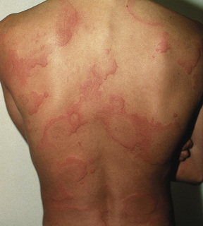
Cené L. Gibson Urticaria, also referred to as hives, is caused by a vascular reaction that occurs in the upper dermis of the skin. It is characterized by the development of wheals on the body surface (Fig. 67-1). Acute urticaria is defined as episodes of hives lasting less than 6 weeks, whereas chronic urticaria is defined as hives persisting for more than 6 weeks. Chronic urticaria can be categorized as idiopathic or autoimmune. On the continuum of urticaria, deeper release of vasoactive mediators into lower dermal and subcutaneous layers of the skin causes angioedema, and severe symptoms can result.1,2 Physical urticaria, which accounts for 15% to 25% of chronic cases,3 is a distinct form of urticaria caused by exposure to physical triggers, such as mechanical or thermal triggers, water, or cold. The hives associated with physical urticaria typically fade within an hour, except in the case of pressure urticaria, in which case the hives take longer to develop and subsequently take longer to fade. In some cases, there is an association between thyroid autoimmunity and urticaria.1 There may be an autoimmune component in some patients with chronic urticaria. An association between Helicobacter pylori and urticaria also has been identified, and recommendations include screening for and/or providing treatment for H. pylori colonization in patients without gastrointestinal complaints.1 Urticaria is a common disorder, affecting an estimated 20% of the population at some time during their lives.3,4 In most cases, it is relatively mild in presentation, albeit frustrating for the patient. In other cases, however, it represents part of a continuum that includes anaphylaxis and can be life-threatening. Acute urticaria is more common in young adults, children, and atopic individuals, affecting 15% to 20% of the general population at some point in their lives.3 This form of urticaria is often attributed to exposure to food allergens, food additives, medications, or radiocontrast media, with rare identification of a specific trigger. Chronic urticaria is slightly more common in middle-aged women and does not show the same predilection for individuals with atopy; 60% of all cases are idiopathic.3,4 Urticaria is an immediate hypersensitivity reaction that occurs after exposure to an allergen or antigen. Mast cells located in the loose connective tissue of the skin release histamine in response to the exposure. Histamine binds to H1 receptors, leading to dilation of capillaries and vascular permeability. Arteriolar dilation leads to flaring around the lesions, and extravasation of fluid from the leaky capillaries leads to wheals, which are superficial itchy swellings in the skin. The histamine that is released causes the pruritus.1,4 Deeper swellings of the skin and alimentary tract can occur in some cases; these swellings tend to be more painful than pruritic and are consistent with angioedema. Histamine also activates H2 receptors, increasing gastric acid secretions.1,4 Mast cells can also be activated by immunoglobulin E (IgE) antibodies stimulated by foods, drugs, insect stings, latex, or animals. Alternatively, activation can occur directly from drugs, including opiates, nonsteroidal anti-inflammatory drugs, and radiocontrast media. Other cell mediators, such as complement and neuropeptides (substance P), may be involved. Recent laboratory research has implicated the complement system in chronic urticaria, particularly C5a, found on cutaneous cells but not on pulmonary cells. This may explain the lack of pulmonary symptoms in chronic urticaria. Certain human leukocyte antigen (HLA) class II associations are also being investigated as possible links to chronic autoimmune urticaria (CAU), and evidence has recently emerged that an underlying autoimmune disease may manifest as CAU.1,4 Patients with urticaria initially note pruritus followed by the development of hives. Lesions appear in crops that last 2 to 3 hours and then disappear, only to flare up elsewhere later. They generally fade in less than 24 hours, leaving no trace. Episodes can occur as often as daily and in chronic urticaria can last up to 2 years. Important history can be simplified into the six Is: infections, ingestants (food), injectants (drugs), insect stings, inhalants (pollen), and internal disease. Latex allergy is an increasingly common cause of urticaria. Other historical factors to be elicited are exposure to heat or cold, fever, exercise, change in menses, and emotional stress. In more severe cases of urticaria, the patient may experience angioedema and may report difficulty breathing. Physical examination reveals edematous pink or red wheals surrounded by a bright red flare. The center of the lesions may be clear or, rarely, may develop bullae. Lesions typically appear on the torso but may occur anywhere on the body. The patient with physical urticaria caused by exposure to some physical stimulus may show what is referred to as dermatographism on examination. Dermatographism is the development of a wheal-and-flare reaction when the skin is stroked with a pen or other physical stimulus. In severe cases of urticaria with angioedema, there may be swelling of the face or oropharynx and deeper swelling in the dermis.
Urticaria
Definition and Epidemiology
 Immediate emergency department referral or physician consultation is indicated for urticaria patients with angioedema, respiratory failure, or hemodynamic compromise.
Pathophysiology
Clinical Presentation
Physical Examination
Related posts:


Stay updated, free articles. Join our Telegram channel

Full access? Get Clinical Tree


