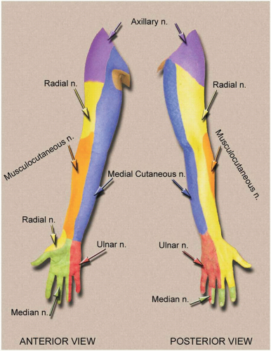
3 trunks: upper (C5, C6), middle (C7), and lower (C8, T1) in the interscalene space, anterior to scalenus medius and posterior to scalenus anterior.
The trunks pass over the lateral border of the first rib and under the clavicle; each trunk (superior, middle, and inferior) divides into anterior and posterior branches. The branches reunite to form cords.
3 cords around the axillary artery:
Lateral cord: lateral portion of the median nerve and musculocutaneous nerve.
Medial cord: medial portion of the median nerve, ulnar nerve, medial cutaneous nerve of the forearm, and medial cutaneous nerve of the arm.
Posterior cord: axillary and radial nerves.
AXILLARY
Cervical C5, C6.
Posterior to the third part of axillary artery.
Run laterally to the surgical neck of the humerus.
Posterior branch gives off the upper lateral nerve of the arm.
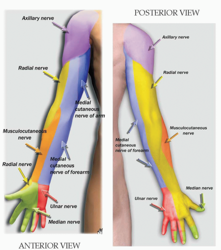
Cutaneous innervation of the shoulder.
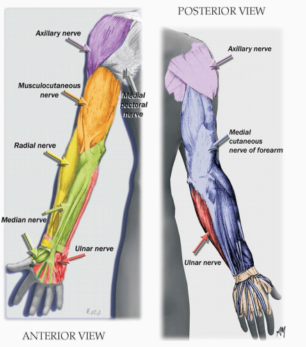
Deltoid.
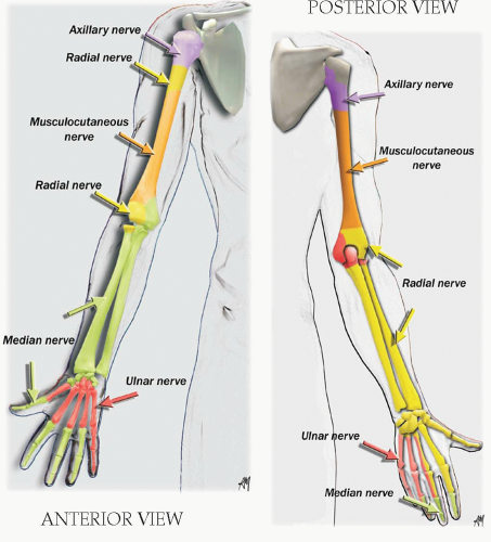
Shoulder joint.
Contraction of the deltoid.
MUSCULOCUTANEOUS
Cervical C5, C6.
Cutaneous innervation of the lateral part of the forearm.
Ventral musculature of the arm.
Flexion of the forearm by contraction of the biceps (long and short head).
Possible confusion with a radial stimulation. The contraction of the supinator and brachioradialis muscles innervated by the radial gives a flexion and supination of the forearm.
The contraction of the supinator and brachioradialis muscles innervated by the radial gives a flexion and supination of the forearm.
RADIAL
Cervical C6, C8.
Posterior to the axillary and brachial arteries.
Run with the profunda brachii artery between the long and medial heads of triceps.
Before leaving the axilla, the radial nerve gives the posterior cutaneous nerve of arm that innervates the posterior upper arm skin.
The posterior cutaneous nerve of forearm innerves the posterior forearm skin.
The deep radial branch innerves the forearm musculature and posterior part of carpal bones.
The superficial radial nerve passes over the tendons of the snuff box and terminates as cutaneous branches to the dorsum of the hand.
Cutaneous innervation of the posterior part of the arm, forearm, and posterolateral part of the hand.
Extensor muscles (dorsal musculature in the upper extremity below the shoulder).
Radioulnar joint and wrist joint.
Extension and supination of forearm.
Extension of the wrist and the fingers.
Abduction and extension of the thumb.
MEDIAN
Cervical C6, thoracic T1.
The median nerve lies lateral to the axillary artery and then lateral to the brachial artery before crossing the artery at the level of the midhumerus.
Gives branch to elbow joint (superior portion of the radioulnar joint): capitulum of humerus, radial head, and epitrochlea of humerus.
Gives branch to pronator muscles at level of forearm (one of the terminal branches is the anterior interosseous n.).
Arises just below the two heads of pronator teres m.
Contraction of flexor digitorum superficialis = flexion of proximal interphalangeal (IP) joints and secondary metacarpophalangeal (MCP) joints and wrist.
Contraction of flexor digitorum profundus (60% to 70% median and 30% to 40% ulnar) = flexion of distal IP joints and secondary flexion of proximal IP and MCP joints and wrist.
Elbow joint (radioulnar joint).
Cutaneous innervation of the lateral palmar skin.
Ventral musculature of the forearm (flexor and pronator muscles).
Wrist and lower radioulnar joint (sensory supply to the anterior part of carpal bones).
Flexion of the fingers and wrist.
Pronation of the wrist.
Opposition of the thumb (adductor pollicis brevis and opponens pollicis).
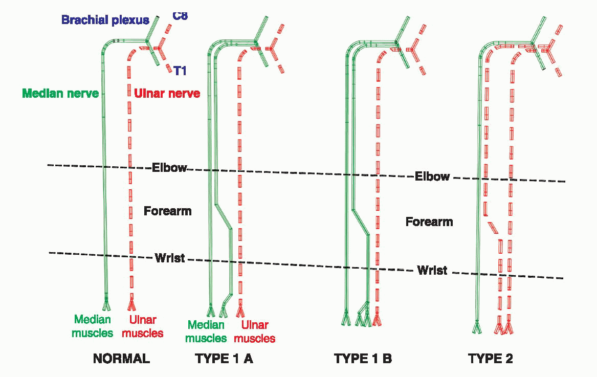
Martin and Gruber Anastomosis
Median and ulnar anastomosis at forearm.
Present in 20% of patients.
Median and ulnar stimulations give the same flexion of the digits (flexion of the last 2 digits and adduction of the thumb) but adduction and flexion of the wrist are exclusively obtained with ulnar stimulation (no anastomosis with flexor carpi ulnaris).
ULNAR
Cervical C8, thoracic T1.
Medial to the axillary and brachial arteries.
Gives a branch to elbow joint: olecranon and medial epicondyle of humerus.
Divides into terminal branches at the pisiform bone.
Dorsal cutaneous branch arises 5 cm proximal to the wrist; passes deep to flexor carpi ulnaris.
Cutaneous innervation of the medial part of the hand.