Lower Extremity Landmarks


Lower Extremity Landmarks


DERMATOMES


MYOTOMES


OSTEOTOMES


SCIATIC NERVE


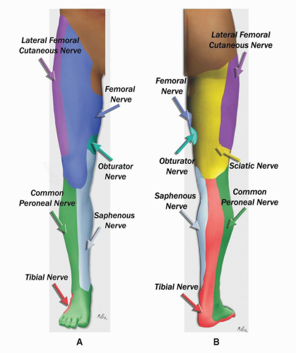
  • Inferior division of lumbar L4, L5 and sacral S1, S2, S3 nerves.


  • Emerges from the greater sciatic foramen.


  • Lies below the piriformis muscle (m.), deep to gluteus maximus m. on the posterior wall of the pelvis.


  • Descends between the greater trochanter of the femur and the ischial tuberosity.


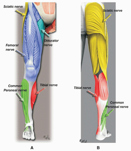
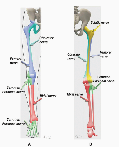
  • Splits into the common peroneal and tibial nerves. This division may take place at any point between the sacral plexus and the lower third of the thigh.


  • Articular branches arise from the upper part of the nerve and supply the hip joint.


POSTERIOR FEMORAL CUTANEOUS NERVE

Sacral S1, S2, S3 nerves.


image


  • Emerges from the pelvis through the greater sciatic foramen below the piriformis.


image


  • All sensory branches.


  • Skin of the perineum.


  • Posterior surface of the thighs and legs.


COMMON PERONEAL NERVE

L4, L5, sacral S1, S2 nerves.


image


  • Winds around the neck of the fibula from posterior to lateral.


  • Divides into superficial and deep peroneal nerves.


  • The superficial peroneal nerve gives innervation to the skin on the dorsum of the foot.


  • The deep peroneal nerve gives innervation to extensor muscles of the ankle and foot and the skin on first dorsal web space.


image


  • Knee joint and ankle joint (deep peroneal).


  • Posterior and lateral aspect of the calf.


  • Tarsal and metatarsal joints.


  • Dorsum of the foot and toes.


image


  • Deep peroneal nerve stimulation.


  • Dorsiflexion of the foot.


  • Eversion.


TIBIAL NERVE

L4, L5, sacral S1, S2, S3 nerves.


image


  • Passes down in the midline into the fossa between the semitendinosus and biceps femoris m. and lies lateral to the popliteal artery.


  • Divides into terminal branches, medial and lateral plantar n., and calcaneal n.


  • Sural n. arises in the poplitea fossa and pierces the deep fascia to become subcutaneous.


image


  • Knee joint and ankle joint (tibial n.).


  • Skin on the lower lateral and posterior part of the calf, the lateral part of the foot, and the little toe (sural n.).


  • Heel and skin of the medial part of the sole (calcaneal n.).


  • Skin of the sole (lateral and medial plantar n.).


image


  • Plantar flexion, inversion.


  • Flexion of the toes.


image


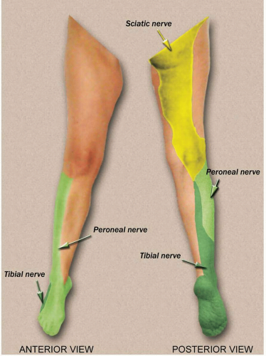
SCIATIC NERVE: DERMATOMES


SCIATIC NERVE BLOCKS

In relation to the lesser trochanter, they are divided into proximal and distal approaches.

In relation to the surface of the thigh, they are divided into anterior, lateral, and posterior approaches.

PROXIMAL POSTERIOR APPROACHES:


  • Parasacral


  • Classic


  • Lithotomy (Raj’ approach)


  • Subgluteal

PROXIMAL LATERAL APPROACH

PROXIMAL ANTERIOR APPROACH

DISTAL APPROACHES:


  • Lateral popliteal


  • High posterior popliteal


  • Classic posterior popliteal


PROXIMAL POSTERIOR APPROACH


PARASACRAL APPROACH

Patient position: Patient in lateral position, side to be blocked being nondependent, both hips and knees flexed.

Landmarks:


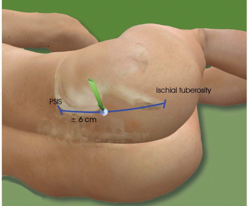
  • Line between the posterior superior iliac spine and the ischial tuberosity.


  • Insertion point is 6 to 7 cm caudal to the posterior superior iliac spine on this line.

Tips:


  • Needle is introduced perpendicular to the skin or at a 30-degree angle in the cranial direction. Upon bone contact (deep landmark), the sciatic nerve is located 2 to 3 cm deeper. This bone contact corresponds to the medial part of the
    greater sciatic notch of the hip bone. The needle needs to be redirected either caudally or laterally or both.


  • A first muscle twitch occurs when the needle is passing through the gluteus muscle. A second, deeper muscle twitch occurs when passing through the piriformis muscle. The nerve is located beneath the piriformis muscle at a depth of 6 to 9 cm.


  • If the first distal twitch is a hamstring contraction, deeper advancement of the needle will result in a tibial nerve stimulation (60%) or a combined tibial and common peroneal stimulation (18%).


  • At this level, the sciatic nerve is close to internal iliac vessels (sciatic vascular trunk).


PARASACRAL APPROACH


CLASSIC POSTERIOR APPROACH

Patient position: Patient in lateral position, side to be blocked being nondependent, with knee and hip flexed (Sim’s position).


Landmarks:


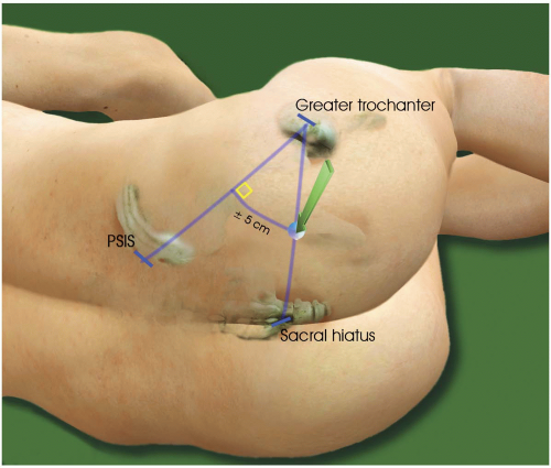
  • Line between the posterior superior iliac spine and the greater trochanter.


  • Perpendicular line is drawn at its midpoint.


  • Intersection with a line between the greater trochanter and the sacral hiatus. Or


  • 5 cm on a perpendicular line drawn at the midpoint of the line between the posterior superior iliac spine and the greater trochanter.

Tips:


  • 4-inch needle.


  • Perpendicular to the skin.


  • The first contraction is elicited when the needle passes through the gluteus maximus muscle; then a deeper muscular contraction occurs when the needle passes through the piriformis muscle.


  • A tibial or peroneal neurostimulation is elicited 1 cm deeper.


  • Multistimulation: a dorsiflexion and eversion of the foot (peroneal nerve) means that the needle is stimulating the lateral part of the sciatic nerve.


    A tibial n. stimulation will be elicited by moving the needle medially.


  • Bone contact = lateral part of the greater sciatic notch of the hip bone. The needle must be redirected medially, caudally, or both.


CLASSIC POSTERIOR APPROACH


LITHOTOMY (RAJ’ APPROACH)

Patient position: Patient in supine position, an assistant holds the leg to be blocked with the knee and hip flexed.

Landmarks: Midpoint of a line between the greater trochanter and ischial tuberosity.

Tips:


  • Needle is introduced perpendicular to the skin.


  • Nerve is located at a depth of 5 to 7 cm.


  • Stimulation of the tibial or common peroneal nerve (hamstrings may be direct muscle stimulation).

Only gold members can continue reading. Log In or Register to continue

Stay updated, free articles. Join our Telegram channel

Jun 5, 2016 | Posted by in ANESTHESIA | Comments Off on Lower Extremity Landmarks

Full access? Get Clinical Tree

Get Clinical Tree app for offline access