Case Presentation
A 47-year-old white man was brought to the emergency department (ED) by ambulance after being found unresponsive by his wife. He was accompanied by empty bottles of amitriptyline and cyclobenzaprine.
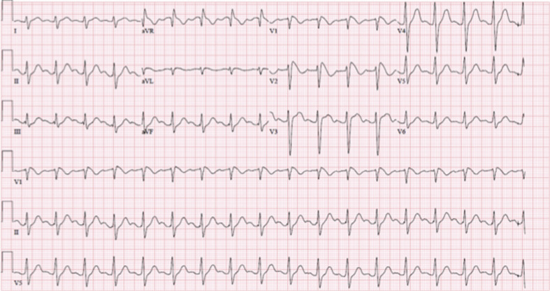
On physical examination, the patient’s vital signs were remarkable for a blood pressure of 94/55 mm HG, pulse rate of 105 beats/min, respiratory rate of 18 breaths/min, and temperature of 35.7°C (96.3°F). Physical examination revealed an obtunded mental status, normal cardiopulmonary examination, and dry skin. Laboratory study results were normal: glucose 117 mg/dL; negative salicylate, acetaminophen, and ethanol concentrations; and a negative urine toxicology screen. An initial ECG ( Figure 1 ) was obtained.

What is the diagnosis?
ECG Findings
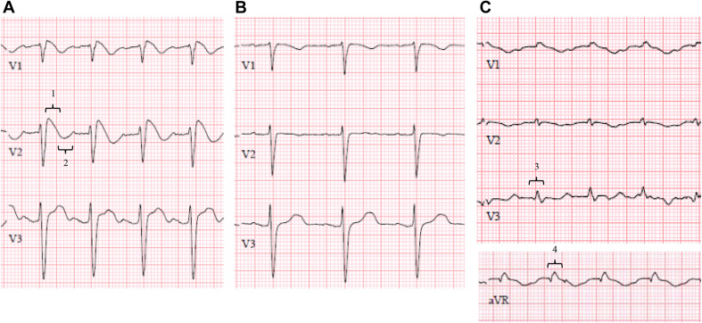
Figure 1 demonstrates sinus tachycardia, right axis deviation, a high-amplitude R wave in aVR, and QRS-interval widening, typical features of tricyclic antidepressant overdose. Figure 1 also demonstrates coved ST-segment elevation in V1 and V2, consistent with a type 1 Brugada ECG pattern (magnified in Figure 2 A ).


