Ultrasound Guided Sciatic Block in the Popliteal Fossa
Colin McCartney
Nick Lo
Applied Anatomy
The sciatic nerve passes into the thigh and lies anterior to the hamstring muscles (semimembranosus, semitendinosus and biceps femoris [long and short heads]), lateral to adductor magnus and posterior and lateral to the popliteal artery and vein (Fig. 41-1). At a variable level (approximately 5 to 10 cm above the popliteal crease) the sciatic nerve divides into the tibial (medial) and common peroneal (lateral) components. The tibial component becomes more superficial as it moves distally accompanying the popliteal vessels in the distal popliteal fossa. The common peroneal nerve moves laterally to a position medial to the tendon of biceps femoris as it moves down posterior and lateral to the knee. Since most foot and ankle surgical procedures involve both tibial and common peroneal components of the nerve it is essential to anesthetize both nerve components. Blockade of the nerve before it divides therefore simplifies the technique.
Block Technique
Prior to the use of ultrasound it was recommended that the needle insertion point was at least 5 cm above the popliteal crease to ensure block of both nerves. However, the use of ultrasound has simplified that process by allowing us to follow the nerves and determine exactly the level of division. Ultrasound therefore allows us to accurately define anatomy prior to needle insertion and to choose a needle insertion point that minimizes distance to the nerve from skin. More importantly, local anesthetic spread can be directed in real time allowing the nerve to be completely surrounded by local anesthetic.
Posterior and lateral ultrasound guided approaches have been reported in the regional anesthesia literature. The sciatic nerve is readily visible on ultrasound deep to biceps femoris and semitendinosus and lies posterior and lateral to the femoral artery (Fig. 41-2). The use of ultrasound removes the need to rely on surface landmarks and the practitioner can choose the most convenient needle insertion point based on the sonographic assessment of the anatomy.
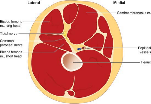 Figure 41-1. Gross anatomy of the popliteal fossa. |
Most often the popliteal fossa is viewed in the transverse plane so that the sciatic nerve appears as a circular structure deep to biceps femoris (Fig. 41-2). The easiest method for finding the sciatic nerve is to follow the tibial nerve, which is a hyperechoic structure lying deep and lateral to the popliteal artery at the popliteal crease, and follow this hyperechoic structure until it is joined further proximal in the popliteal fossa by the peroneal
nerve. The sciatic nerve can be found directly above the popliteal fossa without using this “mapping” technique by looking deep and medial to the biceps femoris muscle and superficial and lateral to the popliteal artery (Fig. 41-2).
nerve. The sciatic nerve can be found directly above the popliteal fossa without using this “mapping” technique by looking deep and medial to the biceps femoris muscle and superficial and lateral to the popliteal artery (Fig. 41-2).
Related posts:


Stay updated, free articles. Join our Telegram channel

Full access? Get Clinical Tree


