Patient Position: Continuous thoracic paravertebral block (TPVB) is performed most easily with the patient in the sitting position with feet dangling over the side of the bed. It is also possible to perform the block in the lateral or prone position.
Needle Size and Catheter: The use of a Tuohy needle is preferred because its blunt, rounded tip provides a distinct “pop” on penetrating the costotransverse ligament and may diminish the chance of perforating parietal pleura. For continuous TPVB use an 18-gauge Tuohy needle with graduated markings and 20-gauge polyamide closed-tip, multiport catheter. More flexible epidural catheters are not recommended as they will be difficult to insert.
Volume and Infusion Rate: An initial injection through the needle of 5 mL of ropivacaine 0.5% to help distend the paravertebral space and facilitate catheter insertion. This is followed by the injection of an additional 10 mL through the catheter. After the surgery, the catheter is infused with 0.2% ropivacaine at a rate of 7 to 10 mL/hr.
Anatomy and Landmarks: The thoracic paravertebral space is a triangular space whose boundaries include anteriorly the parietal pleura, posteriorly the superior costotransverse ligament, and medially the vertebral body, intervertebral disc, and intervertebral neural foramen. The apex of the triangle laterally is continuous with the intercostal space. The space is bisected by the very thin endothoracic fascia which creates two “compartments.” The anterior compartment contains the sympathetic chain, and the posterior compartment contains the intercostal nerve, dorsal ramus, intercostal blood vessels, and rami communicantes. Spinal nerves in the paravertebral space are relatively devoid of fascial covering making them uniquely and exceptionally sensitive to local anesthetic blockade. The important anatomic surface landmark for TPVB is the relevant spinous process or processes for the desired dermatomes to be blocked. Note that the steep angulation of thoracic spinous processes brings them opposite the transverse processes of the adjacent more caudad vertebra.
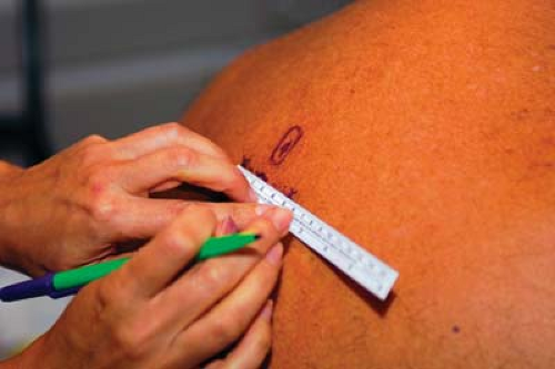
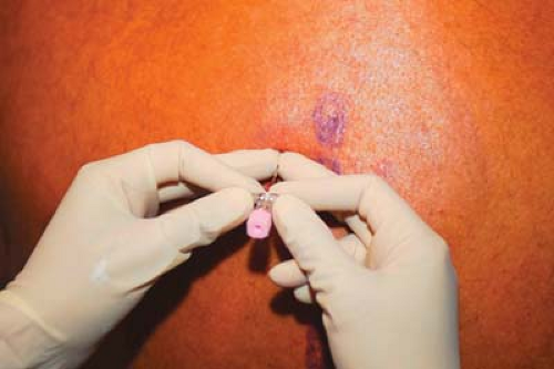
Approach and Technique: The relevant spinous processes are identified and marked on the skin and a point 2.5 cm lateral to the spinous process is also marked (Fig. 31-1). Disinfectant is applied in standard fashion and local anesthetic (1% lidocaine) injected at each injection point using a 1.5-cm 25-gauge needle. The Tuohy needle is advanced onto the transverse process and the depth from skin to paravertebral space marked by placing the index or third fingers on the needle shaft 1 cm from the skin (Figs. 31-2, 31-3). This will now serve as both a depth gauge and a guard against excessive insertion of the needle. The needle is then walked caudally off the inferior border of the transverse process and inserted to a depth 1 cm deeper than the transverse process—that is, to the depth allowed by prior placement of the index fingers on the needle shaft. Typically at this point one feels a confirmatory “pop” upon penetration of the costotransverse ligament. A drop of fluid is placed in the needle hub and the patient is asked to inspire deeply (Fig. 31-4). Correct placement is denoted by lack of movement of the fluid bubble. A drawing inward of the fluid indicates intrapleural needle placement, in which case the needle should be immediately withdrawn. After correct needle placement is thus confirmed, 5 mL of local anesthetic is injected through the needle. It is helpful to have an assistant inject through an extension tube as this helps avoid significant movement of the needle. Following the injection the extension tube is disconnected and the catheter inserted a depth of 3 to 5 cm beyond the tip of the needle (Fig. 31-5). The catheter is affixed in standard fashion using adhesive strips and transparent dressing (Fig. 31-6).
Figure 31-1. The skin is marked at 2.5 cm lateral to the spinous process.
Figure 31-2. The finger placement on the needle shaft will serve as both a depth gauge and a guard against excessive insertion of the needle.
Tips
Depth of the adult paravertebral space from the skin ranges between 3 and 7 cm except for the morbidly obese. Surprisingly, depth correlates only weakly with height, weight, and BMI.
Limit volume of injectate through the needle—the higher pressures when injecting through the needle may force the local anesthetic through the intervertebral foramen into the epidural space.
Any significant resistance to injection through the needle indicates improper needle placement.
Only gold members can continue reading. Log In or Register to continue