Principles of Sonography
Paul Bigeleisen
The frequency of medical ultrasound ranges between 2 MHz and 13 MHz. The average wave length in this band is about 1 mm. This limits the resolution to structures that are larger than 1 mm. Most nerves of interest range in size from 2 mm to 10 mm. Veins and arteries of interest are typically 3 mm to 15 mm.
Many factors contribute to the quality and resolution of the ultrasound image. In general, higher frequency probes generate higher resolution images. Unfortunately, high frequency ultrasound waves (8 MHz to 13 MHz) are rapidly attenuated in tissue so that high frequency probes are best suited for structures less than 5 cm deep to the skin.
The ultrasound beam may be refracted as it passes through tissue. When this occurs, a nerve or other organ may appear at a different anatomical location than its actual site. This is the same phenomenon responsible for the apparent bending of your forearm when you place it in a bucket of water (Fig. 32-1). Fat globules below the skin, in the muscle and around nerves are about 1 mm in diameter. These globules serve as diffraction sites for the incident and reflected ultrasound beam and cause a speckled appearance in the image (Fig. 32-2). Fat is also extremely efficient at absorbing ultrasound so that little of the beam is returned to the receiver. For these reasons, obese patients can be very difficult to image.
The image formed of a nerve on ultrasound is very sensitive to the angle of incidence of the beam relative to the nerve. Sometimes changing the angle of incidence by only a few degrees can bring the nerve into focus. This phenomenon is thought to be caused by diffraction of the type described above.
Modern platforms allow the user to adjust the brightness (gain) of the entire image or more superficial (near field) and deep (far field) structures. Increasing the gain makes the entire image whiter. Increasing the gain too much creates a snowy background whereby all of the structures become indistinguishable. In general, the gain should be set so that most of the background is black and only the structures of interest, such as nerves and vessels, are easily seen. Modern machines also allow the user to adjust the contrast. The formal term for contrast is dynamic range compression. Increasing the dynamic range compression makes the white images whiter and the black images blacker. This may bring the edges of anatomic structure into better view. Decreasing the dynamic range compression makes everything look grey.
Arteries can usually be distinguished by their pulsatile nature. Veins can be distinguished by their compressibility. Pressing on the skin with the probe will usually cause the
vein to collapse. Color flow Doppler imaging can also be used to identify and distinguish arteries and veins. By convention, blood flowing toward the probe is colored red. Blood flowing away from the probe is colored blue. Blood flowing perpendicular to the probe remains black. Velocity gates can be set to measure the flow velocity. High velocities are usually arteries. Low velocities are usually veins.
vein to collapse. Color flow Doppler imaging can also be used to identify and distinguish arteries and veins. By convention, blood flowing toward the probe is colored red. Blood flowing away from the probe is colored blue. Blood flowing perpendicular to the probe remains black. Velocity gates can be set to measure the flow velocity. High velocities are usually arteries. Low velocities are usually veins.
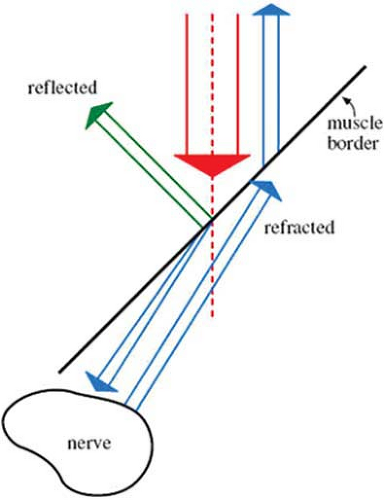 Figure 32-1. Ultrasound beam refracting as it passes through tissue. |
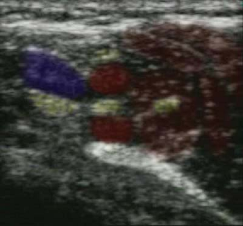 Figure 32-2. Fat globules below the skin serve as diffraction sites for the incident and reflected ultrasound beam and cause a speckled appearance in the image. |
Transducer elements can be arranged in linear or curved arrays (Fig. 32-3). Linear arrays create rectangular images and are most useful for superficial structures. Curved
arrays create wedge-shaped images and are most useful for deeper structures. Because the beam disperses in a curved array, its resolution is usually lower than a linear array. A phased array retains the elements in a straight line. But the elements fire in sequence creating a phase delay between each element. The net result is a wedge-shaped image from a set of linear transducers. Because this signal is averaged, its resolution is also lower than a standard linear array.
arrays create wedge-shaped images and are most useful for deeper structures. Because the beam disperses in a curved array, its resolution is usually lower than a linear array. A phased array retains the elements in a straight line. But the elements fire in sequence creating a phase delay between each element. The net result is a wedge-shaped image from a set of linear transducers. Because this signal is averaged, its resolution is also lower than a standard linear array.
Related posts:


Stay updated, free articles. Join our Telegram channel

Full access? Get Clinical Tree


