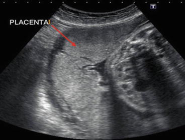
Fig. 43.2
Normal pregnancy ultrasound image
- 1.
- 2.
What risk factors are implicated in this presentation?
- 3.
What is the classification/grading and common sonographic findings?
- 4.
What is the frequency of this presentation?
- 5.
How does coagulation change during pregnancy?
- 6.
What is your choice of anesthesia?
Answers
- 1.
The ultrasound image depicts abnormal placental attachment to the uterine wall, which is characterized by invasion of trophoblast into the uterine myometrium.
- 2.
The incidence of placenta accreta has been increasing and seems to parallel the increasing cesarean section rate. A low-lying placenta (placenta previa) and any condition or surgeries resulting in myometrial tissue damage, along with advanced maternal age and multiparity have been implicated as risk factors.
- 3.
Diagnosis of placenta accreta is usually established by transabdominal and transvaginal ultrasonography and may be supplemented by magnetic resonance imaging (MRI). Abnormal placental attachment is defined according to the depth of myometrial invasion as:
- (a)
Accreta: Chorionic villi attach to the myometrium
- (b)
Increta: Chorionic villi invade into the myometrium
Stay updated, free articles. Join our Telegram channel

Full access? Get Clinical Tree


- (a)


