Fig. 25.1
Technical ABR changes . The first tracing on the top is the baseline while the second set (second from top) was taken during obstruction of the silicone earpiece tube. The lower three traces are taken after the release of obstruction
What was the Cause of This Change? Was it Surgical, Pharmacologic, Physiologic, Positional, or Technical?
A surgical cause could be easily eliminated since the change occurred prior to surgical incision. Changes related to anesthetic agents are usually bilateral. However, because the ABR changes were unilateral (the responses to right ear stimulation were normal), an anesthetic-related cause was unlikely. In addition, short latency ABRs (those measured early in the first 10 ms after stimulation) are generally resilient to anesthetic changes. When changes do occur in the presence of inhalation anesthetic agents, they are limited to small increases in latency that do not exceed 0.75 ms. Nitrous oxide has no effect on ABRs, and narcotics, propofol, and barbiturates minimally affect ABRs [15]. Midlatency ABRs (≥50 ms) are affected by anesthetics, but these were not monitored in this case. The anesthetic agents used for this particular patient had very little effect and could be excluded as the cause of the change.
Physiologic changes related to hypothermia may occur while surgery is in progress—when the brain is exposed to a relatively cool ambient temperature and cold solutions are used for irrigation. The effect of hypothermia has been reported to either increase or decrease the amplitude of the ABR. Some reports state that in the face of hypothermia, latency increases of about 7 % for each 1 °C will affect wave I of the ABR, and below 26 °C this effect will double [15]. This did not occur in this case. Other physiologic factors such as hypotension, hypoxemia, and low arterial CO2 concentration can cause bilateral changes. In this case, there were no such changes in any of these physiologic parameters. In rare situations when a major pathological condition is present in one ear, a physiologic change may lead to bilateral signal changes, with the changes more pronounced on the side with pathology.
We are left with the possibility of either a positional or technical etiology for the encountered change. Because the normal first tracing was done after the patient was positioned with the head fixed without any head or position readjustment, positioning as the cause of the ABR change could be excluded as well.
Technical reasons for ABR changes may include the failure to generate or deliver proper stimulation or the inability to collect and analyze the signals [15]. Disconnected and broken wires and operator errors account for some of the technical changes. Fluid in the ear canal, kinking of the silicone extension tube, or complete or partial dislodgement of an earpiece can all interfere with stimulation intensity and result in decreased amplitude or completely obliterated responses. Placing cotton and wax or ointment over the earpiece protects against such a problem. The inability to properly collect signals may be related to high impedance, broken wires, noise artifacts, and the use of electrical cautery. The use of an ultrasound aspirator or monopolar cautery can saturate the amplifier and lead to poor signal acquisition. In addition, the use of an integer number for the stimulation rate can cause the acquisition system to lock onto rather than to cancel out 60/50 Hz interference commonly associated with the frequency at which power is being delivered and thus interfere with signal acquisition. For more information about technical problems, see Chap. 16, “Wiring and Electrical Interference in Intraoperative Monitoring”.
In this case, a review of the potential technical causes for loss of the ABR response revealed that a near complete kink of the silicone extension tube following draping was the cause. The signals recovered immediately after releasing the kink, allowing the appropriate intensity sound stimulus to be applied.
Case #2
In another scenario similar to Case #1, a 58-year-old man with left-sided disease was placed in the lateral position. The surgeon requested IOM monitoring, to include ABR, cranial nerves 5 and 7 in addition to MEP. Baseline responses revealed normal ABRs on both sides. The MEP baseline responses revealed an absent left hand, but present left foot responses and the presence of both hand and foot responses from the right side (Fig. 25.2).
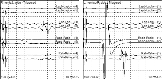
Fig. 25.2
Left panel is tc-MEP from the right hemisphere, showing the missing left hand responses and present left foot response. The right side panel is left tc-MEP with both hand and foot responses
What Could Be the Cause?
In this case we have normal ABRs with absent left hand MEP responses. As in the first case, the surgical procedure had not yet begun, so a surgical cause for the absence of the response was excluded. Anesthesia could be ruled out as a cause since there were no changes in the anesthetic delivery or depth of anesthesia. In addition, changes due to anesthetics are usually bilateral. Physiologic changes such as those due to hypotension, hypothermia, hypocarbia, and hypoxemia are usually bilateral as well. Because there were no changes in any of these parameters, physiologic causes for the absent left hand MEP response were eliminated. However, localized ischemia in the left arm due to a tourniquet is possible, but none had been used. The possibility of a technical cause for the absent response exists. A technical cause would involve either stimulation or recording. Two needles were used for MEP stimulation and because responses were present from the right side and the left lower extremity, stimulation can be excluded as a cause for the absent response. The other possibility to explain this absence is the recording needles in the left hand. These were examined and found to be fine. Low-intensity stimulation is not a possible cause since there were small responses from the other side indicating that the stimulation intensity was strong enough to elicit responses from both sides. In addition, if responses were absent, it would usually be the foot rather than the hand response if the stimulation intensity was too weak. We are left with positioning as the cause of the absent response. Could this be caused by head position and ischemia-related changes? This is unlikely since the ABRs were normal bilaterally and the right side and left foot MEP responses were normal as well. A very localized ischemic event or a stroke is possible but these cannot be diagnosed based on a clinical examination only. A close examination of the left arm revealed that the arm had been stretched backward by the tape used to keep the arm away from the surgical field. Releasing the tape and moving the arm slightly forward resulted in an immediate recovery of the MEP signals. Their absence may have been due to stretching of nerves or ischemia produced by the abnormal position. A pulse oximeter had been placed on the fingers of the right rather than the left hand to monitor for possible ischemia resulting from the lateral positioning, and therefore we were not able to confirm ischemia as the cause of the absent signals. The fast signal recovery points to ischemia as the cause of the absent responses rather than stretching. Left uncorrected, the positioning may have resulted in nerve injury.
Changes in the ABR due to extreme head positions have been reported by Grundy et al. [16]. Severe flexion and twisting of the head resulting from a patient being positioned in the lateral position may alter the relationship of intracranial structures, increase intracranial pressure, decrease cerebral blood flow, and disturb blood flow to the 8th cranial nerve and other areas of the brain, which can result in changes of the ABRs and MEPs. Such changes are generally corrected with readjustment of the head position and restoration of normal blood flow. Inspection of the head position in this case indicated a slight distortion of the head position but major distortion of the arm. After repositioning of the arm to a more neutral position, the signals readily recovered (Fig. 25.3). This patient may also have had abnormal anatomy or some pathology accentuated by the extreme position that led to changes in the MEPs . If the ABR, SSEP, and MEP baselines are obtained after a patient’s positioning is completed and if the signals are severely abnormal, it may be necessary to readjust the position of the head and extremities.
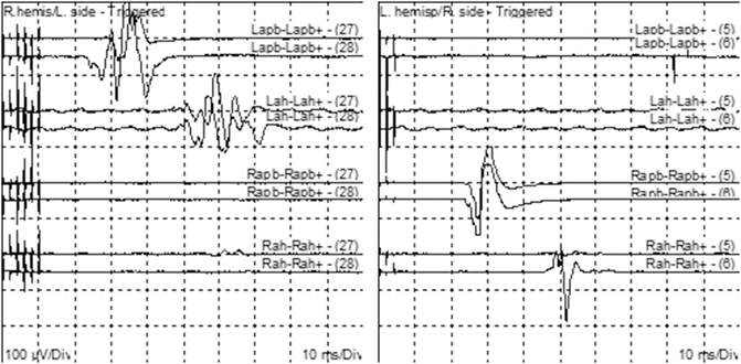
Fig. 25.3
Left side panel is stimulating from right side tc-MEP showing recovery of the left hand responses after adjusting the arm position. The right side is normal responses from left tc-MEP
Case #3
This case is similar to the first case. Baseline values were obtained and the signals were stable before and after positioning of the patient. The surgical procedure began, the dura was opened, and the surgical microscope was used. The surgeon placed and then adjusted a self-retaining surgical retractor several times in order to facilitate surgical exposure and to decompress the trigeminal nerve. During this period, the technologist observed gradual changes of the ABR signals, which included a 5–15 % increase in wave V latency and a 40 % decrease in wave V amplitude from the ipsilateral ear (Fig. 25.4).
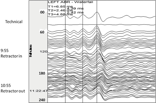
Fig. 25.4
Technical and surgical (retractor ABR changes). The trace on the top is the baseline. Technical change is the second tracing (from top). Cerebellum retractor was placed at 9:55 and released at 10:55. Changes in waves I, III, and V can be seen between these times
Is This a Significant Change?
Polo et al. [17] designated a 0.4-ms (7 %) change in latency of wave V as an early warning sign, a 0.6-ms (10 %) change in latency as the time to alert the surgeon, and a 1-ms (17 %) latency change as serious enough that it needed to be addressed—especially if it was associated with a change in amplitude [17]. In contrast, in 2006, Ramnarayan and Mackenzie [18] found a 0.9-ms latency increase and 50 % amplitude drop to be significant and associated with postoperative hearing loss. Our in-house criteria for alerting the surgeon of a potentially significant change is a progressive 5, 10, and 15 % increase in latency. Some authors depend on latency alone, but Hatayama and Moller [19] believe changes in wave V amplitude are important and should be used as warning criteria when they decrease by 40 %. The changes observed in this case did reach a significant level that required the surgeon’s attention.
What is the Cause of This Change?
Technical and positional causes were excluded, as the patient remained in a stable position and proper functioning of the monitoring system was verified. Both the ambient temperature and the patient’s measured temperature were unchanged, no cold irrigation had been used, and the vital signs were stable. Physiologic causes were therefore unlikely to be the cause of the observed ABR changes . Since the changes were unilateral, pharmacologic- and anesthetic-related causes were doubtful, so surgical factors remained as the likely potential etiology.
Surgical causes of ABR changes can be divided into three categories [20]. The first group includes gradual changes in ABR signals from stimulation of the ear on the operative side that recover with surgical maneuvers such as repositioning of the retractor. These are usually not associated with hearing loss. The second group includes ABR changes where there is an abrupt loss of all waves except wave I on the operative side that do not recover with surgical maneuvers. Such changes are usually associated with postoperative hearing loss. The third group of changes consists of a loss of all ABR signals after wave I on the contralateral side of surgery. This change is usually associated with brainstem dysfunction and may result in severe postoperative neurological deficits beyond just hearing loss.
Surgical causes for ABR changes may be due to mechanical or thermal irritation or injury to the 8th cranial nerve or its blood supply, which result in abrupt loss of the signals [15]. Coagulation of small blood vessels that provide blood flow to the cochlea or of veins that drain from the brainstem may result in such changes. Slower changes in the signals without complete loss may be a consequence of direct compression or traction on the nerve or indirect traction on the nerve through cerebellar retraction and tension on the bridging arachnoid bands. Indirect interference with blood supply to the cochlear nerve caused by compression, coagulation, spasm, or intentional occlusion can lead to varying degrees of ABR changes. Retraction of the cerebellum to improve surgical exposure may lead to stretching and potential damage of the 8th cranial nerve, and be associated with an alteration in ABR signals. Such damage can be more critical in MVD than in cases of tumor resection. During cases of tumor resection, distortion of the nerve by stretch or compression may be better tolerated having occurred over time, whereas during MVD, this anatomical stretch represents an acute insult. Changes related to traction of the cerebellum recover soon, often within minutes after retractor adjustment. If abnormal signals persist, the retractor should be completely withdrawn, and the 8th cranial nerve checked for any evidence of compression including from the Teflon pledgets utilized by the surgeon to isolate the trigeminal nerve from surrounding structures impinging on it.
A clinical situation in which there is wave I delay or loss, with wave V delayed but present, may be the result of mechanical or thermal damage to the cochlea. The presence of wave I associated with equal changes in waves III and V is related to a more proximal injury to the 8th cranial nerve and could be attributed to causes such as cerebellar retraction, nerve compression, or small vessel vasospasm. Preserved waves I and III with delay or loss of wave V may be caused by mechanical, thermal, or vascular injury in the mid to upper pons. Injury to the auditory pathways cephalic to the lower mesencephalon may be associated with normal ABRs. A vascular injury in the posterior fossa that spares the auditory pathway can be associated with normal ABRs.
Related posts:
 Deep Brain Stimulation
 Intraoperative Neurophysiologic Monitoring During Surgery for Supratentorial Mass Lesions
 Surgery for Chiari Type I Malformation
 Intraoperative Neurophysiological Monitoring for Intracranial Aneurysm Surgery
 Intraoperative Monitoring in Tethered Cord Surgery
 IOM Instrumentation Layout and Electrical Interference
Stay updated, free articles. Join our Telegram channel

Full access? Get Clinical Tree


