Age (year)
HR/min
RR/min
Newborn
100–180
30–60
0.5
80–160
25–40
1–5
80–130
20–30
6
75–115
18–25
8
70–110
18–25
10
70–110
15–20
12
F 70–110, M 65–105
12–20
14
F 65–105, M 60–100
12–20
16
F 60–100, M 55–95
12–20
18
F 55–95, M 50–90
12–20
Table 18.2
Pediatric normal values: blood pressure (BP) at rest
Age (year) | BP (at rest) 90th percentile (systolic/diastolic) | |
|---|---|---|
F | M | |
Newborn | 76/68 | 87/68 |
0.5 | 106/65 | 105/66 |
1 | 105/67 | 105/69 |
2 | 105/69 | 106/68 |
4 | 107/69 | 108/69 |
6 | 111/70 | 111/70 |
8 | 114/72 | 114/73 |
10 | 117/75 | 117/75 |
12 | 122/78 | 121/77 |
14 | 125/81 | 126/78 |
16 | 127/81 | 131/81 |
18 | 127/80 | 136/84 |
Psychosocial Considerations
The emotional impact on children, parents, and society cannot be neglected. The initial evaluation and management of children in the trauma setting can often be traumatizing to both the staff and parents. Proper counseling, debriefing, and post-hospital care are essential when addressing the pediatric trauma patient. It is equally important to consider prevention strategies and effective injury prevention and control require a comprehensive and integrated system that can monitor and collect essential data as the ongoing data collection systems recommended by WHO [15].
Pediatric Management Challenges
The previously mentioned peculiarities of the pediatric trauma patient may lead to challenges in executing appropriate management in a timely fashion in this special population. Establishing initial differential diagnoses of the patient’s injuries depends to a great degree on an appropriate history and physical examination as well as pertinent laboratory and radiologic investigations. Obtaining an accurate and comprehensive history from pediatric patients can be a challenge due to patients’ inability to communicate or due to events that are unwitnessed. Collateral history from parents, when available, can be impaired by the stress and emotional impact of the situation on the parents [16–18]. Certain signs may raise the suspicion of child abuse [19, 20]; subsequently a new set of rules should be considered in obtaining the history as recently recommended by the American Academy of Pediatrics [21] that may include interviewing and examining the child without the parents’ presence. The physical exam of a pediatric trauma patient can vary significantly in reliability based on the experience of the examiner. That involves simple tasks such as assessing volume status [22] or even measuring the temperature [23]. For instance, reading chest X-ray in emergency room for children with suspected pneumonia is reported to have high degree of intra-observer variability [24]. More advanced investigation can be limited in children due to the invasive nature of the test, the need for sedation, the increased risk of radiation exposure, or the inability to get consent in a short time. Because of concerns over ionizing radiation in this age group ultrasound carries a particular value, as will be explained later. Reducing diagnostic errors in pediatric emergency department can be achieved by adopting some of the key principles such as the ones suggested by Croskerry and others [25–27]. Croskerry, who is specialized in Emergency Medicine, hypothesized that the diagnostic process in ED follows the so-called dual process theory. The theory suggests that two basic modes of thinking are in action to reach a diagnosis. Type 1 processes are fast, reflexive, and intuitive, and may operate at a subconscious level. In contrast, Type 2 processes are analytic, slow, and deliberate that require focused attention. Using checklists [28] with better utilization of system 2 (Table 18.3) and de-biasing and disengagement of the intuitive mode (Table 18.4) are among the key concepts [29] to avoid diagnostic errors in the ED and in decision making in general.
Table 18.3
Hierarchical de-biasing strategies
Universal | Critical thinking training |
Dual process theory training | |
Cognitive/affective bias training | |
Generic | Structured data acquisition |
Get more information | |
Be more skeptical | |
Slow down/reflection | |
Rule out worst-case scenario (ROWS) | |
Consider the opposite | |
Specific | Bias inoculation strategies |
Re-biasing strategy | |
Specific forcing functions | |
Stopping rules | |
Checklists |
Table 18.4
Enhancement of system 2
Reflective, careful thinking |
Evidence of critical, analytical, skeptical, and disciplined thought |
Willingness to look beyond the obvious |
Seeks out and considers alternate explanations |
Looks for counter evidence |
Absence of obvious cognitive or affective bias, stereotyping |
Sound logical reasoning |
Intellectual honesty |
Evidence of critical thinking in analysis of clinical scenarios |
Ability to distinguish between weak and strong evidence |
Different types of pediatric shock require fast diagnosis and management. Obstructive shock can be due to severe pulmonary hypertension that may occur as a response surge of inflammatory mediators typically in sepsis, massive blood transfusion, severe hypoxia, or acute lung injury. Pulmonary vasodilators should be considered in these cases. Common causes of obstructive shock can be diagnosed by bedside focused ECHO, particularly pericardial tamponade. The distributive shock due to sepsis progresses rapidly in pediatric patients, usually with peripheral vasoconstriction and poor perfusion and far less common (compared to adults) with peripheral vasodilation, or the so-called warm sepsis [30–32]. Therefore, the use of vasodilators (e.g., phentolamine) with inotropes (e.g., epinephrine) or milrinone may constitute a consideration in these cases [33]. Appropriate and prompt correction of glucose (hypoglycemia), acidosis (hydrogen ion abnormalities), and hypoxia is considered among the top “5 Hs” priorities in shocked/arrested child [34]. Calcium correction is particularly important in the pediatric population. In goal-directed therapy of pediatric shock, care should be given to calcium that interacts with many physiological functions particularly myocardial contraction and inotropy [35, 36]. Ca2+ facilitates both systolic contraction (inotropy) and diastolic relaxation (lusitropy). Depletion of Ca2+ stores, impaired uptake of Ca2+ by the sarcoplasmic reticulum (SR), or impairment of the Ca2+-regulating proteins, including SR-Ca2+-ATPase, sarcoplasmic-endoplasmic reticulum Ca2+ (SERCA), and phospholamban, can contribute to rapid deterioration of cardiac contraction and thus aggravate the shock state. Treatment of acidosis and other metabolic abnormalities require rapid correct diagnosis that can be facilitated by following a few rules in the interpretation of the blood gas. Table 18.5 provides ten rules of thumb for such conditions and can be used as a pocket card and is available as smartphone image for quick reference. As in adults, multiple doses of etomidate should be used with caution in children with shock conditions because of its potential effects on the hypothalamic–pituitary–adrenal axis [37].
Table 18.5
Ten rules for acid/base balance interpretation
Respiratory acidosis/alkalosis (pH and PCO2 opposite direction) | ||
Rule 1 | The rule of (1–4 and 2–5) | |
Respiratory acidosis (1–4) | Each rise of PCO2 by 10 mmHg will be compensated by increase of bicarb by 1 in acute and 4 in chronic | |
Respiratory alkalosis (2–5) | Each drop of PCO2 by 10 mmHg will be compensated by drop of bicarb by 2 in acute and 5 in chronic | |
Rule 2: | Each acute change Δ of PCO2 by 10 mmHg will Δ pH by 0.08 | |
Metabolic alkalosis (pH and PCO2 same direction) | ||
Rule 3 | PCO2 = 0.9 × HCO3 + 15 | |
Rule 4 | HCO3 + 15 = last 2 digits of pH (7.xy) | |
Rule 5 | Metabolic alkalosis can be | |
Type | Chloride resistant | Chloride resistant |
Urine Cl < 10 mmol/L | Urine Cl > 20 mmol/L | |
Causes | Volume depletion | K or Kg depletion |
Vomiting | High mineralocorticoids (aldosterone) | |
Villous adenoma | High glucocorticoid (cortisol) | |
Diuretics | Excess alkali (bicarbonate-citrate) | |
Drugs | Drugs (antacid-effervescent) | |
Treatment | Treat the VVVDD | Correct K and Mg |
Normal saline (especially if chloride is low) | Treat the cause | |
Drugs | ||
Acetazolamide or ammonium chloride | ||
or hydrochloric acid | ||
Metabolic acidosis (pH and PCO2 same direction) | ||
Rule 6 | ||
(Rule 6.1) | Anion gap (AG) = Na − (CI + bicarb) | |
Normal = 12, AG > 19 then it is primary metabolic acidosis | ||
(Rule 6.2) | gap/gap ratio = Δ AG: Δ,bicarb = (AG-12}:(24-bicarb) | |
If it is =1 then high AG acidosis | ||
If it is <1 then high AG + normal AG acidosis | ||
If it is >1 then mixed metabolic acidosis + alkalosis | ||
(Rule 6.3) | Metabolic acidosis can be either | |
(a) High AG acidosis | (b) Normal AG acidosis | (c) Pseudo norm AG acidosis |
Added acid | Loss of alkali | False low AG |
Lactic acid | Drainage (biliary/pancreatic) | Low albumin |
Uric acid | Dilution | Paraprotiens |
Acetic acid | Diamox | Lipids |
Ketoacid | Diuretics | High Ca-High Mg |
Amino acid | Disease | Polymyxin B |
Salicylic acid | (Addison/renal tubular acidosis) | Lithium poisoning |
Rule 7 | Rule of 7 each drop of pH by 0.1, the PCO2 will drop by 7 mmHg to compensate | |
So if | pH | PCO 2 |
7.40 | 35 | |
7.30 | 28 | |
7.20 | 21 | |
7.10 | 14 | |
Rule 8 | Any Δ in pH by 0.01 (not due to PCO2) is due to base Δ by 0.67 | |
Rule 9 | Total body bicarb deficit = base deficit × body Wt. × 0.3 | |
Rule 10 | (Interpretation steps) | |
Identify the primary abnormality | ||
See PCO2 and pH directions | ||
Calculate expected compensation and compare to the actual value | ||
Look for the mixed abnormality | ||
Integrate with clinical data | ||
Remember, the body NEVER overcompensates | ||
“Critical” Ultrasound in the Pediatric Patient
The published reports [38–44] about the FAST (focused assessment with sonography in trauma) exam in children carried conflicting results but in general indicate low sensitivity. The physiological and anatomical differences that are explained earlier in this chapter may explain the difference in the FAST utility in children versus adults and may also explain the relatively less uptake of FAST in pediatric population. Despite the fact that a negative FAST exam does not have a useful rule-out value, a positive FAST exam is still very useful in stratifying the management of pediatric trauma cases.
Beyond the ultrasound role in the initial detection of abdominal fluid by FAST, ultrasound (US) recently gained an increasing role in stabilization of shocked patient in the ED. A new philosophy of US imaging recently emerged which is the “problem-based critical” ultrasound. This is a paradigm shift from being an organ-based, systematic, comprehensive diagnostic exam to a new concept of problem-based, goal-directed, focused multi-organ, time-dependent exam done by the treating physician. Based on this concept, the US exam is not just done for detection of fluid collection or for an organ or region (e.g., US for abdomen or chest) but rather done to enhance the management of a problem (US for hypotension, US for hypoxia, US for sepsis, etc.). Certain problem-based US exams can involve scanning of multiple organs to enhance the management of a certain patient’s problem. The US within this concept is not considered just as a diagnostic tool but rather as an integral part of ED management that involves diagnosis, tailored treatment, monitoring effects, goal-directed therapy, ruling out possible complications, and of course guiding most of the pediatric ED procedures [45]. If we consider the example of “ultrasound for hypotension,” a process of scanning of three organs (heart, inferior vena cava (IVC), and lung) will generate a “sonodymanic” pattern to enhance the management of hypotension as follows [46]:
1.
Gross evaluation of right and left ventricular functions. This is usually done without detailed quantitative measurements but rather done by global qualitative exam of both right ventricle (RV) and left ventricle (LV). This quick exam also rules out/in any pericardial collection.
2.
Vena cava analysis. A small IVC with spontaneous collapse suggests hypovolemia (flat IVC) that is usually fluid responsive. A distended IVC (fat IVC) will be fluid non-responsive and may suggest hypervolemia, pulmonary hypertension, or tamponade. When interpreted with pertinent echocardiographic signs (step 1), then the diagnosis can be immediately established [46]. In addition to IVC, also superior vena cava and aortic flow have been used to assess fluid responsiveness in children and adults [46]. However, because of the variability in pediatric body size and growth, absolute values are not helpful and only relative indices [47–49] such as the ones listed in Table 18.6 may have potential application in the pediatric patient. This however remains an area waiting for validation by better quality studies in the pediatric trauma population. In the presence of a fixed and distended IVC, a small and hyperkinetic right ventricle is suggestive of tamponade (especially if combined with rapid accumulation of pericardial fluid) with possible right ventricle or right atrial collapse. On the other hand, a dilated and hypokinetic right ventricle may signify pulmonary hypertension.
Table 18.6
Relative indices for ultrasound assessment of fluid responsiveness
Name | Symbol | Equation | Cut valuea (%) | R# |
|---|---|---|---|---|
IVC collapsibility index | Δ IVC | 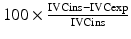 | >18a | 47 |
SVC collapsibility index | Δ SVC | 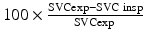 | >36 | 48 |
Ao velocity index | Δ AoV | 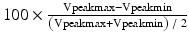 | >12 | 49 |
3.
Get Clinical Tree app for offline access

Assessment of the lung. A profile of lung ultrasound is characterized by multiple horizontal artifacts (A-lines) which are findings of well-aerated normal lung. With the loss of air in the lung and development of different lung pathologies, several artifacts and new ultrasound features may appear (as summarized in Table 18.7 and Figs. 18.1, 18.2, 18.3, and 18.4), particularly the appearance of abnormal multiple vertical lines (B-lines). A hypokinetic LV and a B-line profile by lung ultrasound in a hypotensive pediatric patient with low oxygen saturation are suggestive of cardiogenic pulmonary edema, indicating thus the administration of diuretics and inotropes. Once this is suspected, fluid administration in such cases should be extremely cautious. That is part of “ultrasound for hypoxia” that includes the lung and heart exam. The presence of an A-line profile in lung ultrasound usually rules out pulmonary edema. In addition to heart assessment, simple ultrasound signs (Table 18.8) for the lung can differentiate cardiogenic from non-cardiogenic pulmonary edema [50]. Some reports claim the ability of ultrasound to differentiate between bacterial and viral pneumonia in pediatric ED with using the six-zone lung scanning technique which is somewhat different from the adult approach [51].
Table 18.7


Key points in lung ultrasound
Related posts:


Stay updated, free articles. Join our Telegram channel
Full access? Get Clinical Tree