Parameter
Change in pregnancy
Heart rate
Baseline rate increased by 15 bpm
Blood pressure (systolic and diastolic)
Nadir of 15 mmHg below baseline by the end of the second trimester
Cardiac output
Increased by 1–1.5 L per minute over baseline
Total blood volume
Increased by 50 % above normal
Red blood cell mass
Lesser increase compared to plasma volume resulting in dilutional anemia (lower hematocrit)
Respiration
Increased tidal volume and minute ventilation, with resultant hypocapnea
Lung volumes
Decreased residual volume (elevation of diaphragm)
Gastrointestinal motility
Delayed gastric emptying
Gastrointestinal anatomy
Displacement of bowel into upper abdomen
Musculoskeletal
Widening of symphysis pubis and sacroiliac joints
Fibrinogen
Increased (normal fibrinogen may hail the development of disseminated intravascular coagulation)
Pseudocholinesterase levels
Decreased—consider lower dose of succinylcholine during rapid sequence induction
Hematocrit
32–42 %
White blood cell count
5,000–12,000/μL
Arterial pH
7.40–7.45
Bicarbonate
17–22 mEq/L
PaCO2
25–30 mmHg
Fibrinogen
>3.79 g/L (3rd trimester)
Shock in the Pregnant Patient
Due to the physiologic increase in intravascular volume, hemorrhage may initially be well compensated for in the pregnant trauma patient. It is typically only after significant blood loss that tachycardia and hypotension are encountered. At that point, however, catecholamine-induced maternal shunting of blood away from the uterus and placental vasculature will have deprived the fetus of vital perfusion resulting in compromise. Maternal shock is associated with an 80 % fetal mortality rate [23]. Abnormal fetal lie (oblique or transverse), easy palpation of fetal parts, and/or inability to palpate the uterine fundus may suggest uterine rupture, while placental abruption is associated with vaginal bleeding, uterine tetany, and severe abdominal pain. Unfortunately, these clues to suggest obstetrical causes of hemorrhage are not sufficiently sensitive (e.g., vaginal bleeding is absent in 30 % of cases of placental abruption). An additional vital sign is therefore required in the assessment of shock in the pregnant trauma patient: the fetal heart rate (see Fetal Monitoring). Early fetal assessment with frequent intermittent or continuous fetal monitoring should be performed during maternal stabilization, as fetal heart rate changes may be the first indicators of maternal hypovolemia and impending shock. When using this approach, not only will resuscitation of the mother be improved, but the risk of fetal complications, including fetal demise, will be decreased.
Distinct Aspects of the Pregnant Trauma Patient
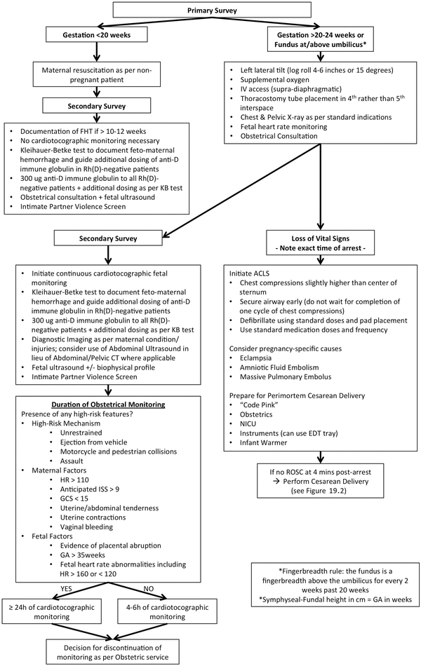
While the management of thoracic and abdominal injuries during pregnancy differs little from the nonpregnant state, there are certain considerations unique to the pregnant trauma patient. All female trauma patients should be stratified as either potentially pregnant, pregnant at less than 23-weeks gestation, pregnant at greater than 23-weeks gestation, or peri-mortem. Fetal viability is achieved at approximately 23-weeks gestation, at which time maternal resuscitation and fetal assessment should take place concurrently. Gestational age (GA) and the estimated date of delivery (EDD) —or “due date”—is determined using the date of the last normal menstrual period (LMP) (Naegele’s rule: EDD = LMP − 3 months + 7 days) or a “dating” ultrasound performed at 8–12-weeks gestation [24]. In order to err on the side of fetal viability, if an accurate gestational age is not immediately known at the time of the patient’s presentation, physical examination may be used as a surrogate indicator for an estimated GA of at least 20–24 weeks (Fig. 19.1). Initial evaluation of a reproductive-age woman should include a concise and focused obstetric and gynecologic history, with universal pregnancy testing performed [22]. In some instances, early pregnancy may be diagnosed during surgeon-performed ultrasound [25].


Fig. 19.1
Assessment and management of the pregnant trauma patient. Based on the content in Katz, 2012 [65]. ACLS advanced cardiac life support, CT computed tomography, EDT emergency department thoracotomy, FHT fetal heart tones, GA gestational age, GCS Glasgow coma score, HR heart rate, ISS injury severity score, NICU neonatal intensive care unit, ROSC return of spontaneous circulation
A Kleihauer-Betke (KB) test should be performed in all pregnant trauma patients, regardless of Rh status, to identify significant feto-maternal haemorrhage and guide anti-D immune globulin treatment in Rh(D)-negative women [26, 27]. All pregnant Rh(D)-negative trauma patients should receive anti-D immune globulin therapy due to the risk of feto-maternal hemorrhage and subsequent alloimmunization. A standard dose of 300 μg is given, with additional doses pending results of the KB test, where the extent of feto-maternal hemorrhage is quantified. The blood bank should be consulted for dose calculation, timing, and route of administration. There is no relationship between the severity of maternal injury and the incidence of feto-maternal hemorrhage [17, 26], but a positive KB test has been associated with subsequent preterm labor after maternal trauma with the extent of feto-maternal hemorrhage correlating with the likelihood of preterm labor [26]. Fetal complications of feto-maternal hemorrhage include neonatal anemia, cardiac arrhythmias, and fetal death [13]. In cases of significant feto-maternal hemorrhage, fetal intrauterine transfusion may be required.
Preeclampsia is defined as hypertension with proteinuria or adverse conditions (Table 19.2) [28]. Complications of preeclampsia may result in traumatic injuries, such as a motor vehicle collision secondary to visual impairment. Differentiating symptoms of preeclampsia from those of traumatic injury may therefore be difficult in such situations. An eclamptic seizure may mimic traumatic brain injury. Abdominal pain and placental abruption may result from preeclampsia or sustained blunt trauma. Coagulopathy may be caused by preeclampsia-induced disseminated intravascular coagulation or a consumptive process secondary to ongoing traumatic hemorrhage. In each scenario the correct diagnosis is required to tailor and guide treatment.
Symptoms and signs |
|---|
Maternal symptoms |
Persistent or new/unusual headache |
Visual disturbances |
Persistent abdominal or right upper quadrant pain |
Severe nausea or vomiting |
Chest pain or dyspnea |
Maternal signs |
Eclampsia (seizure) |
Severe hypertension (systolic BP ≥ 160 mmHg) |
Pulmonary edema |
Placental abruption |
Abnormal maternal laboratory testing: |
Elevated serum creatinine, AST, ALT, or LDH |
Platelet count <100 × 109/L |
Serum albumin <20 g/L |
Fetal morbidity |
Oligohydramnios |
Intrauterine growth restriction |
Absent or reversed end-diastolic flow in the umbilical artery by Doppler velocimetry |
Intrauterine fetal death |
A search should be made for conditions unique to the injured pregnant patient, such as blunt or penetrating uterine trauma, placental abruption, preterm labor, or premature rupture of membranes. Preterm labor complicates up to 5 % of maternal traumas [17] and may be treated with tocolytics. Corticosteroids such as betamethasone or dexamethasone should be administered to patients between 23- and 34-week gestation in the setting of preterm labor or premature rupture of membranes in order to promote fetal lung maturity [29, 30]. However, delivery should not be delayed to achieve corticosteroid benefit if obstetrically indicated (e.g., significant placental abruption, fetal distress).
A qualified surgeon and obstetrician should be consulted early in the evaluation of a pregnant trauma patient. In the setting of blunt trauma, splenic and retroperitoneal injuries are more common in pregnancy because of increased vascularity [13, 31], and pelvic fractures have been reported to cause direct fetal skull fractures or intracranial injuries [32–34]. In the setting of penetrating trauma, a trauma surgeon should be involved for consideration of surgical exploration to rule out bowel or diaphragmatic laceration [12, 35]. The pattern of anticipated injuries is modified by pregnancy, with upper abdominal penetrating injuries more likely to result in bowel injury due to cephalad displacement from the gravid uterus, versus decreased visceral and retroperitoneal injury with lower abdominal penetrating injuries (but higher uterine and fetal injuries in such cases), where more non-operative management may be considered [35]. Diagnostic peritoneal lavage, if performed, should employ the open technique, above the uterine fundus [36]. Exploratory laparotomy is typically well tolerated and preferable to delayed diagnosis of intra-abdominal injuries, but is associated with an increased risk of preterm labor [37]. Excessive uterine manipulation and maternal hypotension must be avoided. Careful consideration should be given to the need for cesarean section, with the determination of fetal age, fetal maturity, and fetal well-being factored in the decision making. Cesarean section will increase the intraoperative blood loss and operative time, while delayed recognition of fetal distress will increase the risk of perinatal loss [38]. Hysterotomy in the setting of an otherwise uninjured uterus may be necessary for adequate abdominal exploration or repair of identified injuries [16, 39]. If not required at the time of laparotomy, hysterotomy may be avoided in cases of fetal demise where induction of labor may be preferred [39].
Formal uterine and pelvic examination should be performed by the obstetrical team, if available, to evaluate for uterine contractions or tenderness, vaginal bleeding, rupture of membranes, or cervical dilation in addition to evaluation for vaginal lacerations in the setting of pelvic fracture. Amniotic fluid can be identified by its alkaline pH and characteristic ferning pattern on microscopy. Cardiotocographic monitoring must be skillfully interpreted for fetal heart rate abnormalities. Venous thromboembolism prophylaxis should be provided in the form of low-molecular-weight heparin as per standard therapy. Obstetrical follow-up should be arranged to ensure identification and management of late complications, since adverse obstetrical outcomes such as preterm delivery and low birth weight are increased in trauma patients [5, 40]. Screening for intimate partner violence is essential (Table 19.3).
1. Have you been kicked, hit, punched, or otherwise hurt by someone within the past year? If so, by whom? |
2. Do you feel safe in your current relationship? |
3. Is there a partner from a previous relationship who is making you feel unsafe now? |
Fetal Monitoring
Continuous cardiotocographic fetal monitoring should be performed in all viable pregnancies (i.e., beyond 20–24-week gestation), since maternal hemodynamic parameters are not accurate predictors of fetal distress or risk of fetal loss [38, 41]. Cardiotocography will identify signs of placental abruption, fetal distress, and uterine contractions if monitored by a team member with experience in fetal heart rate interpretation. Care must be taken to distinguish the maternal versus fetal heart rate during monitoring. Continuous tachycardia, bradycardia, or repetitive decelerations may be indications for urgent delivery. Monitoring should continue throughout maternal resuscitation and continue for a minimum of 4–6 h in the absence of risk factors for fetal loss in an awake, asymptomatic patient [13, 42], versus ≥24 h in the presence of risk factors or altered level of consciousness (Fig. 19.1) [43] since the risk of immediate complications are increased in these patients [5]. Uterine rupture is strongly correlated with fetal mortality, the signs of which may be missed during assessment of the mother alone [44]. Monitoring should be performed regardless of the apparent low severity and location of injuries sustained by the mother, as fetal compromise has been identified in up to 20 % of cases of apparent minor injury [41, 43]. Placental abruption has been reported in 3 % of cases of minor abdominal injuries and 40 % of cases of severe blunt abdominal trauma where fetal mortality may be as high as 60 % [44, 45]. Similarly, the symptoms of placental abruption may be minimal, depending on the location of retro-placental bleeding and the degree of placental detachment. Even ultrasound may fail to identify placental abruption in 50 % of cases [46, 47]. Such adverse outcomes, however, are expected to manifest within 4–6 h of presentation [43]. In the presence of a reassuring fetal heart rate pattern for 4–6 h, in the absence of uterine contractions (<6 contractions per hour) at a gestational age of >20 weeks, the incidence of late fetal or maternal complications has been found to be minimal and safe discharge following maternal clearance is supported in appropriately selected patients [44].
Radiology
The risk of fetal radiation exposure must be balanced with the benefit of radiographic investigations and/or image-guided procedures. It is impossible to reap the benefits of minimized radiation exposure unless one is alive to do so. It should be emphasized that the best initial treatment for the fetus is the provision of optimal resuscitation of the mother.
Imaging and investigations—including the use of iodinated contrast material—necessary to facilitate diagnosis and management of the injured pregnant patient are endorsed in guidelines published by the American College of Obstetricians and Gynecologists [48] and the American College of Radiology [49]. Consideration should be given to the degree of fetal radiation exposure in the selection of imaging modalities where a choice exists, and abdominal-pelvic lead shielding used where appropriate. Radiation levels should be kept as low as reasonably achievable and the use of specific dose-reduction techniques should be applied [50]. A medical physicist should be involved to calculate the actual fetal dose received, with prospective fetal dose estimation through placement of dosimeters on the patient at the level of the uterus a useful adjunct [50].
Fetal risks of anomalies, growth restriction, or spontaneous abortions are not increased with radiation exposure of <5 rad (1 rad = 10 mGy (milligray)) [48]. Table 19.4 lists estimated fetal exposure levels from common radiologic procedures. When tabulated using this data, a diagnostic imaging work-up that includes chest and pelvic plain radiographs, extremity radiographs, and a “pan-scan” (CT of the head, cervical spine, single-phase CT chest/abdomen/pelvis including thoracolumbar spine) would not necessarily exceed the threshold of 5 rad. Indeed, Tien et al. [51] examined actual delivered radiation doses in trauma patients admitted to a Canadian Level 1 trauma center and found a mean dose of 2.27 rad with an average of 4.9 CT scans and 13.7 plain radiographs performed during their hospital stay. However, some modalities may expose the fetus to doses of radiation that exceed the safe limit, such as pelvic angiography and embolization in the setting of pelvic fracture. Such patients should be counseled appropriately by the obstetrical or perinatology service. The teratogenicity of radiation in pregnancy is most pronounced in the first trimester, highlighting the need for universal pregnancy testing in women of reproductive age [22, 52]. In addition, while no in vivo tests in animals have demonstrated mutagenic or teratogenic effects with low-osmolality contrast agents there are no good studies in pregnant women, and there are theoretical concerns of hypothyroidism developing in the newborn infants [53]. Therefore, the American College of Radiology suggests that these agents should ideally be used in pregnancy only if (a) the information cannot be obtained without contrast administration, (b) the information will affect the care of the patient during pregnancy, and (c) the exam is urgent and cannot be delayed until after pregnancy [53]. Obviously most trauma imaging would satisfy these criteria.
Table 19.4
Fetal absorbed dose from selected radiographic examinations
Procedure | Fetal absorbed dose | ||
|---|---|---|---|
ACOG No. 299 [48] | McCollough et al. [84] | Wieseler et al. [50] | |
Plain radiographs | |||
Chest (AP and lateral) | 0.02–0.07 mrad | 0.2 mrad | – |
Abdominal (single view) | 100 mrad | 100–300 mrad | – |
Extremities | – | <0.1 mrad | – |
Cervical spine (AP and lateral) | – | <0.1 mrad | – |
Thoracic spine (AP and lateral) | – | 0.3 mrad | – |
Lumbar spine (AP and lateral) | – | 100 mrad | – |
CT scans (single acquisition) | 64-row multidetector | ||
Head | <1,000 mrad | 0 | – |
Chest (routine and for PE) | <1,000 mrad | 20 mrad | 2 mrad |
Abdomen and lumbar spine | 3,500 mrad | – | – |
Abdomen | – | 400 mrad | 130 mrad |
Abdomen and pelvis | – | 2,500 mrad | 1,300 mrad |
Abdomen for stones (kidneys, ureters, and bladder) | – | 1,000 mrad | 1,100 mrad |
Angiography | – | – | 1,300 mrad |
Angiography of aorta (chest through pelvis) | – | 3,400 mrad | – |
CT pelvimetry | 250 mrad | – | – |
Background fetal dose for 9 months of pregnancy | – | 50–100 mrad | – |


