Fig. 53.1
Normal AV in ME AV SAX view (a and b), severely stenotic AV in ME AV SAX (c) and ME AV LAX views (d), color Doppler (e) of severely stenotic AV showing turbulent flow at the aortic valve and root. Aortic sclerosis (f)
- 2.
Diagnosis and assessment of severity of AS made on the basis of history, physical exam, and echocardiographic findings. Patients with severe aortic stenosis (AS) usually present with angina, syncope, sudden death, or heart failure. Physical exam may reveal a crescendo-decrescendo ejection murmur. ECG will show signs of left ventricular hypertrophy and left atrial enlargement.
Two-dimensional echocardiography with Doppler evaluation (TTE or TEE) is the test of choice to confirm the diagnosis of AS and assess severity and also note the presence of coexisting diseases such as aortic regurgitation, mitral stenosis, mitral regurgitation, aortic root dilation, and coronary artery disease (Fig. 53.1).
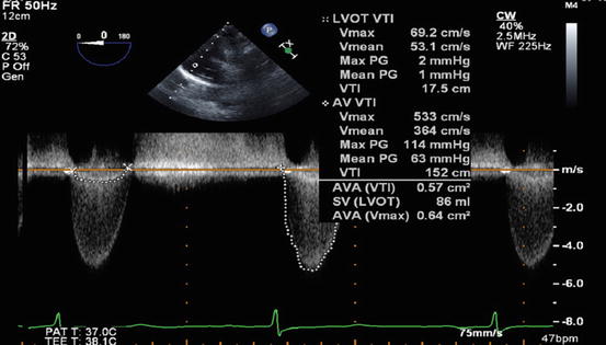
Fig. 53.2
Continous wave interrogation of stenosed aortic valve with measurement of maximum and mean pressure gradient and transvalvular velocity across aortic valve
The peak velocity and mean pressure gradient across the aortic valve are measured by means of Doppler interrogation of the aortic valve (Fig. 53.2). Accurate Doppler measurement of aortic valve velocity (and pressure) requires a near parallel alignment of the ultrasound beam to the aortic valve. The normal aortic valve area is approximately 3.0–4.0 cm2. The velocity and pressure gradients across the aortic valve are flow dependent. In patients with low ejection fraction, dobutamine or exercise stress echo may be needed to confirm the diagnosis. In the 2014 guidelines, severity of AS was divided into 4 stages (A, B, C, and D) based on valve anatomy, valve hemodynamics, hemodynamic consequence, and symptoms [1] (Table 53.1).
Table 53.1
Stages of valvular aortic stenosis (Adapted from the Nishimura et al., 2014 ACC/AHA Practice Guidelines for the Management of Patients with Valvular Heart Disease)
Stage | Symptoms | Valve anatomy and hemodynamics |
|---|---|---|
A | Asymptomatic but at risk | Bicuspid AV Aortic sclerosis (vmax < 2 m/s) |
B | Asymptomatic but progressive | Mild-moderate leaflet calcification, some reduction in systolic motion (vmax 2–3.9 m/s, Pmean < 20–39 mmHg) |
C | Asymptomatic severe AS | |
C1 | Asymptomatic severe AS | Severe leaflet calcification or stenosis with severely reduced leaflet opening (vmax ≥ 4 m/s; Pmean ≥ 40 mmHg, AVA ≤ 1 cm2) |
C2
Related posts:Stay updated, free articles. Join our Telegram channel
Full access? Get Clinical Tree
 Get Clinical Tree app for offline access

| ||

