Fig. 24.1
MRI of 64-year-old woman that shows a large tumor mass in the right cerebellopontine angle (CPA). The cerebellum is shifted to the left hemisphere and the brainstem is compressed
Anesthesia management. General anesthesia was induced using continuous infusion of remifentanil (0.4 μg/kg/min), a bolus of 20 mg etomidate and 35 mg rocuronium.
Total intravenous anesthesia (TIVA) was used to maintain general anesthesia (infusion of 5 mg/kg/h propofol (83 μg/kg/min) and 0.4 μg/kg/min remifentanil). Additional neuromuscular blockade (NMB) was avoided because of intended monitoring of the corticobulbar tract (pathway of cranial nerve VII).
The level of hypnosis was monitored using the EEG-based bispectral index (BIS) recorded from the left forehead. A transesophageal Doppler probe was inserted after positioning of the patient to detect air embolism.
Potential Problems and Structures at Risk
Brain surgery in the sitting position bears the risk of air embolism. In addition, sufficient perfusion pressure must be maintained.
The sitting position has some potential benefits. It allows a better exposure of the surgical field, improved drainage of CSF, reduced blood loss, less tissue damage, reduced ICP, and decreased rate of cranial nerve damage. Potential risks of the sitting position include postoperative quadriplegia, damage to peripheral or cranial nerves, postoperative pneumocephalus, and venous or paradoxical air emboli.
Sudden alterations of the cardiovascular and respiratory systems may occur during or after brainstem manipulation with surgery in the posterior fossa or as a result of the patient’s pathology due to the tight space. The size of the posterior fossa is limited and a small increase of infratentorial volume may produce high pressure on the brainstem with dramatic changes in the vital signs and mental status. This may produce sudden apnea without previous warning signs, e.g., changes in alertness or consciousness. Isolated pathology in the posterior fossa will not lead to dilatation of the pupils. Preoperatively, posterior fossa tumors are associated with the risk of hydrocephalus, damage to cranial nerves, and pressure to the brainstem. Intraoperative manipulations of the brainstem may induce sudden changes in heart rate, blood pressure and arrhythmia (pons, roots of cranial nerve V (trigeminus), IX (glossopharyngeus), and X (vagus)), or bradycardia (periventricular gray, reticular formation). Postoperative complications can occur as sequelae of damage to the cranial nerves. Damage to the trigeminal nerve (V) may lead to lesions of the cornea and damage to the facial nerve (VII) may lead to exsiccation of the eye. Impairment of the vestibulocochlear nerve (VIII) may induce postoperative dizziness and hearing loss and injury to the caudal brain nerves (IX, X, XII) impairs swallowing and increases the risk of aspiration.
Postoperative complications may occur due to swelling or bleeding and can be manifested as sudden apnea or alterations in mental status. Hyperperfusion or clotted blood vessels, leakage of CSF or disruption of CSF circulation, and pneumocephalus are typical postoperative complications.
Monitoring
The goals of hemodynamic monitoring are to ensure adequate central nervous system perfusion, maintain cardiorespiratory stability, and detect and treat air embolism. Blood pressure should be measured at the head level to ensure adequate cerebral perfusion pressure while central venous pressure is measured at the heart level. For monitoring air embolism, transesophageal echo (TEE) is the most sensitive method of detection. However, precordial Doppler is the most widely used method.
Neurophysiological Monitoring
- A.
Positioning of the patient in the sitting position requires flexion of the head and may lead to a dramatic decrease in the perfusion of the cervical spine. This is due to different perfusion zones, especially of the posterior spinal arteries at the cervical and upper thoracic levels. Since the dorsal columns are served by the posterior cervical arteries, monitoring of the somatosensory tract via SSEPs is useful for detecting regional ischemia. Figure 24.2a, b shows recordings of the SSEPs after stimulation of the left and right median nerves during positioning of the patient’s head. Only a few minutes after flexion, the amplitudes of the cervical (C7 level) and cortical responses from both sides were dramatically reduced. Following correction of the head position, all the SSEP responses recovered. Some teams also monitor motor-evoked potentials to assess ischemia in the motor tracts with positioning.
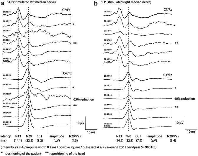
Fig. 24.2
Cervical and cortical SSEP recordings after stimulation of the left (a) and right (b) median nerve during positioning of the patient in the sitting position. Anteflexion of the head (*) led to dramatic depression of cervical and cortical amplitudes. Recovery of all responses after repositioning of the head (**). No remarkable change of latencies was found
A global reduction in the amplitude of both cervical and cortical responses may also be due to a global change of the patient physiology or systematic technical errors and such causes should be ruled out. Appropriate stimulation can be verified by assessing the evoked response at the level of the Erb’s point. Appropriate positions and contact impedances of the recording electrodes must be verified. General anesthesia should be maintained at as steady a state as possible to minimize amplitude changes as a result of the anesthetic agents. In this case, there was no SSEP recording at the Erb’s point and therefore a systematic technical error cannot be excluded (e.g., electrical disturbance of the reference electrode). Therefore, the electrodes were rechecked. With bilateral stimulation and symmetric changes, an error at the stimulus level is very unlikely. There was no change of the continuous propofol infusion and no bolus doses were given.
- B.
The placement of retractors for approaching the tumor can cause shifting of cerebellar tissue which can either involve direct compression of the vestibulocochlear nerve (CN VIII) or indirect impairment of the brainstem. Auditory brainstem responses (ABRs) provide excellent information about the integrity of the auditory pathway and are sensitive to ischemia or tissue damage to the brainstem.
Ipsilateral recordings of ABRs after stimulation of the right and left ear are shown in Fig. 24.3a, b. In contrast to ABRs acquired on the left, the initial recordings from the right side show unstable waves I–III. In addition, the latency of the IV/V complex and the interpeak latencies of I–V were prolonged on the right side (Table 24.1). It is likely that the large size of the tumor and chronic compression of the brainstem led to instability of particular waves and prolongation of the conduction time.
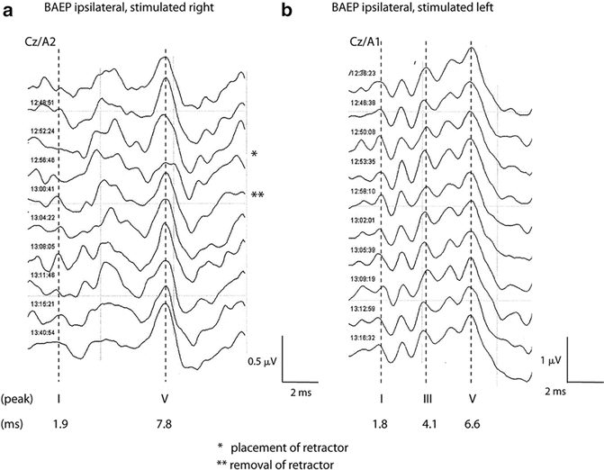
Fig. 24.3
Recordings of ipsilateral ABRs after stimulation of the right (a) and left (b) ear monitoring the episode of retractor placement. Notice the initially prolonged latency of peak V in the right (tumor side) and the unfavorable signal-to-noise ratio with unstable recordings of peak I and III. Peak V is reproducible and stable. Immediately after positioning the retractor (*), the amplitude of peak V (right) dropped about 40 %. The surgeon was informed by the neurophysiologist and with the removal of the retractor (**), the response completely recovered. In contrast, no change was observed in the left-sided response
Table 24.1
ABR ipsilateral recordings


