Structure and Development of the Upper Respiratory System in Infants and Children

Chapter 35 Structure and Development of the Upper Respiratory System in Infants and Children


Respiration is vital to survival and is the first function that is protected in any critical medical situation. The respiratory tract can be divided into the upper or conducting airways and the lower or gaseous exchange airways. For the purposes of this chapter, the trachea down to the carina is included as part of the “upper” or conducting airways. The upper airway shares its development with that of the upper digestive tract, and the lower airway shares its development with the cardiovascular system.


The upper airway serves numerous functions, including air conduction, warming, purification, humidification, protection of the lower airways, and phonation, which is most often overlooked in the critical care setting.


In the fetus, the larynx allows fetal breathing (of amniotic fluid) until the moment of birth, when it assumes the role of protecting gas exchange between the environment and the newborn. Protection of the lower airway and phonation occur simultaneously. Thus the infant’s first cry heralds its ability to sustain independent life. After breathing, eating becomes the infant’s next priority, requiring some of the most complex neurologic coordination found in nature. When confronted with the challenges of maintaining a patent airway, deglutition, and vocalization, a well-functioning upper airway is of utmost importance.


Developmental Anatomy of the Upper Airway


The embryologic development of the nasal cavity, mouth, nasopharynx, oropharynx, and hypopharynx occurs in a separate developmental environment from that of the larynx, trachea, bronchi, and lung parenchyma. Early errors in development can affect both anatomic areas, but these errors are usually incompatible with life. Because the nose, mouth, pharynx, and part of the larynx develop from different embryonic structures than the rest of the larynx and lower respiratory tract, there are few coincident congenital anomalies between these two contiguous but developmentally distinct areas.


The branchial arches, which begin to appear during the fourth week of embryogenesis, give rise to most of the upper airway—nasal passages, pharynx, larynx, and striated muscles— involved in breathing and swallowing. The development of these structures is usually complete by week 14.


The respiratory system, including parts of the larynx, the trachea, and the lungs, also begins to appear during week 14, when the laryngotracheal groove develops into a diverticulum that subsequently separates from the pharynx. In the fourth and fifth weeks, the longitudinal tracheoesophageal folds fuse, forming the tracheoesophageal septum and dividing the foregut into ventral and dorsal portions. The ventral portion becomes the larynx, trachea, bronchi, and lungs, and the dorsal portion becomes the esophagus.


The embryogenesis of the larynx is complex. The cartilages and muscles are derived from the fourth and sixth branchial arches, and the epithelium is derived from the endoderm of the laryngotracheal tube. As this epithelium rapidly proliferates, the larynx is temporarily occluded until the 10th week, when recanalization occurs. Failure to recanalize can result in laryngeal webs, stenosis, or, rarely, atresia. The epiglottis forms by mesenchymal proliferation of the third and fourth branchial arches (Figure 35-1).
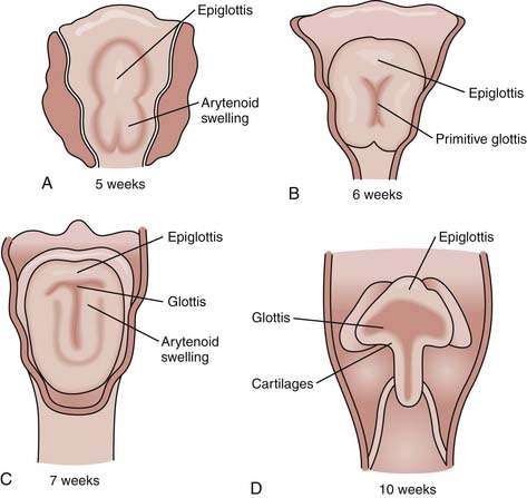


The tracheobronchial tree also has several embryonic origins. Its epithelium is derived from the laryngotracheal tube, and its connective tissue, cartilages, and muscles are derived from the surrounding splanchnic mesenchyme. All the cartilages of the trachea are C-shaped and thus are incomplete posteriorly, giving the airway flexibility to expand, except for the cricoid cartilage immediately below the true vocal folds. The cricoid cartilage is anatomically considered part of the larynx. It is the only cartilage in the airway to form a complete ring. Because of its inflexibility, edema from intubation and inflammation can result in serious, often avoidable injury.


Anatomy and Physiology of the Upper Airway


Nasal Passages


The upper airways begin at the tip of the nose and the vermilion border of the lips. Both the nasal and oral passages allow air stream from the environment through the larynx into the lungs where oxygen and carbon dioxide are exchanged through the alveoli into the cardiovascular system via capillaries. The mouth, oral cavity, and pharynx (oropharynx and hypopharynx) have the additional function of ingesting adequate amounts of food for growth and development.


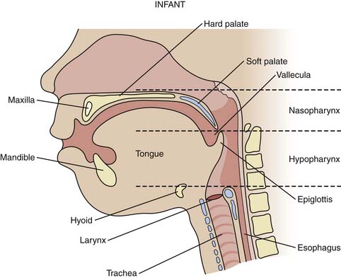
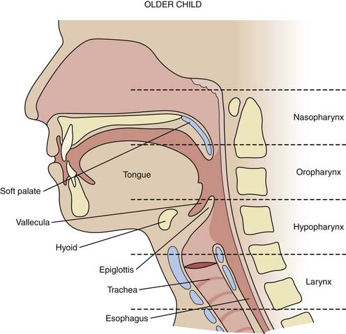
The structure of the upper airways differs in the infant (Figure 35-2), young child, and young adult (Figure 35-3). Preferential nasal breathing is present in neonates and persists up until 6 months of age due to the high riding larynx in the neck with the soft palate and vallecula in close anatomic approximation.


< div class='tao-gold-member'>

Stay updated, free articles. Join our Telegram channel

Jul 7, 2016 | Posted by in CRITICAL CARE | Comments Off on Structure and Development of the Upper Respiratory System in Infants and Children

Full access? Get Clinical Tree

Get Clinical Tree app for offline access