Fig. 36.1.
(a) T2-weighted axial oblique image of the pelvis demonstrating a cleft sign (arrow) just inferior to the pubic symphysis (PS); (b) T2-weighted sagittal image of the pelvis demonstrating hyperintense aponeurotic tears (arrows) of the rectus abdominis (RA) and adductor longus (AL) tendon insertions into the pubic bone (P).
Diagnosis
The history, physical exam, and radiologic findings were consistent with the diagnosis of a sports hernia with adductor tendonitis. Other differential diagnoses should include iliopsoas strains or bursitis, avulsion injuries of the pubic bone, nerve entrapment syndromes, stress fractures of the femoral neck or pubic rami, vertebral body pathology, and associated hip injuries [1]. The most common hip pathology associated with a sports hernia is a labral tear, which is diagnosed with MRI [2]. Other diagnoses that are not part of the musculoskeletal system include appendicitis, urinary tract infections, testicular pain, varicoceles, round ligament entrapment, endometriosis, and ovarian cysts.
Nonoperative Management
Nonoperative management is initially provided for adductor tendonitis associated with groin pain and may include 3–9 months of rehabilitation with a physical therapist. These modalities may involve extensive stretching, deep tissue massage, electrical stimulation, and local injections. These injections employ mixtures of steroids with local anesthetics, and are injected directly into the pubic symphysis, pubic tubercle, or associated aponeuroses. Mixtures commonly include methylprednisolone mixed with 0.5 % bupivacaine , but other combinations of methylprednisolone, dexamethasone, lidocaine, and bupivacaine have been used as well. Overall, the majority of adductor tendon injuries should resolve with inactivity and nonoperative therapy. The goal of nonoperative therapy should restore a full range of motion, while maintaining muscle strength and preventing contracture of the adductor tendon or rectus sheath. Ultimately, the patient should regain strength, flexibility, and endurance quickly if nonoperative management is successful. However, chronic injuries located along the musculotendinous junction usually require operative intervention if athletic activities cannot be halted.
Operative Management
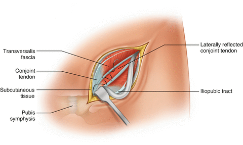
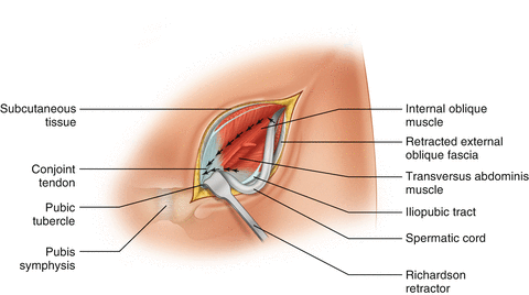
Our technique entails an inguinal incision along the skin crease located directly above the superficial ring. The dissection extends through Scarpa’s fascia to the external oblique aponeurosis. Tears in the external oblique aponeurosis are repaired with 4-0 nonabsorbable sutures since these tears may cause nerve impingements. The external oblique aponeurosis is then opened superiorly and inferiorly through the superficial ring. The spermatic cord is dissected at the level of the pubic tubercle, retracted laterally, and skeletonized to exclude a hernia sac. Once a hernia sac is excluded, a relaxing incision is made along the fascia of the conjoined tendon starting at the level of the deep ring, and extended inferiorly along the pubic bone (Fig. 36.2). The anterior rectus sheath is then released from the pubic bone by incising the aponeurosis approximately one cm superior to the pubic bone. This relaxing incision is then extended laterally until the transversalis fascia of the inguinal floor is encountered. The undersurface of the relaxing incision is undermined in an avascular plane. The sports hernia repair is performed by plicating the previously released fascia of the conjoined tendon to the iliopubic tract with a continuous 2-0 Prolene suture (Fig. 36.3). The first layer starts at the pubic tubercle and extends superiorly to the deep ring. A second layer of 2-0 Prolene imbricates the initial layer. At this point, 10 cc of 0.25 % bupivacaine is injected into the pubic tubercle. Mesh is not placed since the intent of the repair is to lateralize the vector force away from the pubic symphysis and tubercle.


Fig. 36.2.
Relaxing incision starts at the pubic tubercle and extends along the conjoined tendon to the level of the deep ring. The relaxing incision also extends medially along the conjoined tendon to release the aponeurosis from the pubic symphysis for approximately 2 cm.

Fig. 36.3.


After undermining the conjoined tendon beneath the relaxing the incision, the flap is secured with a running 2-0 Prolene suture to the iliopubic tract. A second 2-0 Prolene is used to imbricate the first suture line.
Stay updated, free articles. Join our Telegram channel

Full access? Get Clinical Tree


