Spinal Injury

Chapter 101


Spinal Injury image


The intensivist managing the patient with major trauma in the intensive care unit (ICU) setting must be vigilant for the presence of a coexistent injury to neural or osteoligamentous components of the spine. The history and neurologic examination determine the functional status and level of the neurologic injury, whereas imaging studies determine the integrity and stability of the osteoligamentous complex. Despite the advances of imaging investigations, plain films remain of value for defining instability, especially in the cervical spine. Computer-assisted imaging (computed tomography [CT]) and magnetic resonance imaging (MRI) are complementary studies, each of which contributes valuable information to the assessment and management of the patient with a spinal injury (Table 101.1).


Three goals guide the ICU management of patients with spinal injuries: (1) preventing neurologic injury or progression of an existing neurologic deficit, (2) enhancing the physiologic environment in which neurologic recovery takes place, and (3) stabilizing the spinal column. This chapter presents the management principles and the pharmacologic, nonsurgical, and surgical interventions used to achieve these goals.


Pathophysiology and Biomechanics of Spinal Injury


Spinal cord injury can be subdivided into primary and secondary injures. In the setting of trauma, the primary injury results from the application of force to the spinal cord, causing vascular injury or direct injury to the neuronal and non-neuronal cell populations. The severity of the primary spinal cord injury remains the strongest predictor of neurologic outcome.


Secondary spinal cord injury results from a cascade of physiologic and biochemical events that follow the primary injury. The cascade involves the formation of oxygen-free radicals, cell membrane disruption, and cell death. Factors such as ischemia and hypoxia can accelerate local metabolic injury, emphasizing the need for rigid control of systemic blood pressure and oxygenation in the management of a patient with a spinal cord injury.


The cervical spine (C-spine) consists of the atlantoaxial complex and the subaxial C-spine. The major articulation at the atlantoaxial complex involves the odontoid process and the anterior arch of C1, which is stabilized by the transverse ligament. Direct ligamentous injury and bony injuries that cause incompetence of the transverse ligament complex produce atlantoaxial instability. These include some C1 fractures, most odontoid fractures, transverse ligament injuries, and complex atlantoaxial fractures. Unstable subaxial C-spine injuries are diagnosed on lateral C-spine films obtained in a neutral position and during flexion and extension.


Unstable injuries of the thoracic spine are more likely to occur at the thoracolumbar junction or in the lumbar spine because of the stabilizing capabilities of the rib cage and sternum.


Assessment of Spinal Cord Injury


The first priority with respect to managing any critically injured patient remains assessment and stabilization of the airway, breathing, and circulation. Unnecessary manipulation of the patient’s spine, however, should be avoided before radiographic confirmation of stability.


Inspection of the body for superficial abrasions and contusions can assist in the differential diagnosis of the neurologic injury, localizing it either centrally or to the periphery. Palpation of the entire spine may provoke pain, which can assist in localization of the level of a significant spinal injury. Pain, however, does not determine the extent and stability of the spinal injury. Any pain identified by manual examination requires imaging of that segment of the spine to assess for an osteoligamentous injury. On occasion, with extreme spinal trauma, widening of the interspinous space or step-offs between adjacent vertebrae can be appreciated by palpating posteriorly. Such findings occur rarely in isolation and patients with such findings often have severe neurologic deficit, localized pain, or both.


The primary purpose of the neurologic examination is to assess the integrity of peripheral neural function. A limited assessment of cognitive and cranial nerve function should be performed to determine the presence of intoxication, hypothermia, or brain injury as these processes can interfere with the interpretation of the examination of the peripheral nervous system. The presence of a unilateral neurologic deficit, except in the instance of penetrating spinal injury, is more typical of an intracranial abnormality or involvement of a peripheral nerve, or nerve root, or plexus. Acceleration and deceleration injuries of the spinal cord generally produce symmetric deficits.


A complete assessment of spinal cord function necessitates a thorough examination of sensory, motor, and reflex function. In particular, a segment-by-segment assessment of each dermatome and myotome must be made to determine the level and completeness of the spinal cord injury.


Sensory Examination


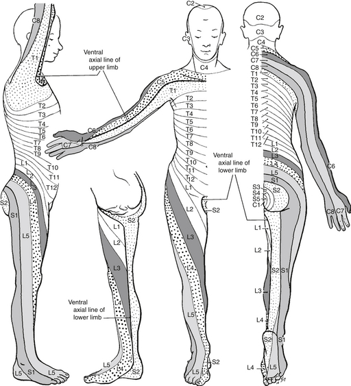
The sensory examination should include assessment of both pain perception and proprioception as these two modalities travel through distinct anatomic tracts in the spinal cord (spinothalamic tracts and posterior columns, respectively). Appreciation of noxious stimulus requires a dermatome-by-dermatome assessment from the highest cervical to the lower sacral levels (Figure 101.1).


The lower cervical and upper thoracic dermatomes (C5-T2) are not represented on the anterior torso. The upper cervical dermatomes (C3-4) extend to the supramammary region, immediately superior to the T3-4 dermatome. Examination of the torso alone results in a sensory assessment that makes the transition from the C4 to the T4 levels, not directly testing the intervening dermatomes. For this reason, a detailed examination must be carried out in the arms and hands to assess these areas. Otherwise, a patient with a low cervical injury can be misdiagnosed as having an upper thoracic injury by a sensory assessment limited to the torso.


The upper extremity sensory assessment must include all six dermatomes represented on the arm for adequate localization of a deficit. Individual leg dermatomes should also be assessed, although disparity in sensory function in a dermatomal pattern after a spinal cord injury is less common in the legs (see Figure 101.1).


The sacral dermatomes, located within the perineal region, should also be tested (see Figure 101.1). The presence of perineal sensation alone (“sacral sparing”) in a patient who otherwise has no demonstrable neurologic function represents an incomplete spinal cord injury, which may have a better prognosis for recovery of spinal cord function than can be expected in a patient with a complete injury.


The assessment of posterior column function involves position and vibration sense testing. Because pain and proprioceptive fibers travel within the same peripheral sensory nerves, posterior column testing can involve the distal aspects of the upper and lower extremities alone, after the detailed noxious stimulus assessment. A disparity in the results of sensory assessments between proprioceptive and pain appreciation occurs only in a patient with a partial cord injury affecting either the posterior or anterior aspects of the cord alone. Transverse injury, or an injury involving a peripheral nerve, should affect spinothalamic and posterior tracts with equal severity.


Reflex Examination


The reflex examination includes deep and superficial reflex assessments. The deep tendon reflexes are tested in the arms and legs. The significance of abnormal reflexes depends on the location and time course of the injury.


In the presence of an acute complete spinal cord injury, the deep tendon reflexes below the level of the lesion are hypoactive. In some instances, normal reflex activity may be seen in the hyperacute state. Hyperactive reflexes develop in the subacute phase (4 to 6 weeks) after an injury because of the loss of inhibition from descending corticospinal pathways. The Babinski reflexes follow the time course of the deep tendon reflexes. In the acute phase of an injury to the spinal cord, they remain unreactive, or a flexor response is occasionally identified. Extensor plantar response develops from chronic compression of the spinal cord or in the subacute phase of an injury.


In acute trauma, the differential diagnosis of hypoactive reflexes includes nerve root injury, plexus injury, and spinal shock. The underlying cause of hyporeflexia is determined by the pattern of the patient’s neurologic symptoms and associated motor and sensory deficits. In addition, hypoactive reflexes may be due to a preexisting condition such as peripheral neuropathy or chemotherapy use, or they may be a normal variant.


Focal hypoactive reflexes in the upper extremities are often the result of a nerve root or brachial plexus abnormality. If the motor and sensory deficits correspond to multiple, adjacent motor or sensory root levels, a plexus injury is likely the underlying cause. Contusions or abrasions of the skin, or underlying fractures involving the shoulder girdle structures, pelvis, or transverse processes, can be associated with these deficits, and their presence should be sought to confirm the diagnosis.


Spinal shock is a phenomenon that is present after a complete spinal cord injury. The patient has absence of all volitional and reflex neurologic activity below the level of the lesion. In contrast, neurogenic shock is a hemodynamic phenomenon. After spinal cord injury at or above the T5 vertebral level, patients may become hypotensive. Characteristically, patients have a relative bradycardia in the presence of low blood pressure. The shock results from the loss of sympathetic outflow. This causes peripheral vasodilation and pooling of blood, which, in turn, impairs venous return to the heart. Loss of sympathetic outflow also has negative inotropic and chronotropic effects on the heart.


Superficial reflexes are diminished in the presence of a spinal cord injury. Nerves from T6-9 and T10-12 subserve the upper and lower superficial abdominal reflexes, respectively. The absence of both upper and lower superficial abdominal reflexes indicates that the lesion is above T6. The absence of lower and preservation of upper superficial abdominal reflexes indicates that the lesion is below T9. Likewise, the L1-2 segmental nerves mediate the cremasteric reflex, and loss of this reflex is pathologic.


The tone of the anal sphincter should also be assessed as part of a full neurologic workup. It is usually diminished or absent in the presence of a complete acute spinal cord injury.

< div class='tao-gold-member'>

Stay updated, free articles. Join our Telegram channel

Jul 7, 2016 | Posted by in CRITICAL CARE | Comments Off on Spinal Injury

Full access? Get Clinical Tree

Get Clinical Tree app for offline access