Fig. 27.1
ABR responses with changes due to retractor effect
Differential Diagnosis of ABR Findings
Several nonphysiological processes may contribute to changes in ABRs during IOM [14]. Inherent to any IOM, ABRs are susceptible to mechanical failure and operative errors such as malfunctioning equipment, accidental removal of the stimulating earpiece, or an obstruction in the tubing connected to the earpiece. These conditions may artificially produce increases in latency, decreases in amplitude, or even an entire loss of signal recordings. Other possible nonphysiological processes that can affect ABR signal recording include artifact created by electrocautery or ultrasonic aspirating devices. In the present case, all the data collection and recording equipment was inspected and determined to be working properly. The use of electrocautery was infrequent and no cavitational ultrasonic surgical aspirator devices were being utilized. We therefore determined that changes in ABR recordings were not caused by a nonphysiological etiology.
Short-latency ABR recordings are relatively resistant to the effects of intravenous and volatile anesthetic agents even at levels of anesthesia that attenuate or abolish other forms of IOM (EEG, SSEP, etc.). However, ABRs become more susceptible to anesthetic agent levels when the patient is hypothermic or hypotensive. While reversible prolongation of peak latencies have been observed in patients undergoing induced hypothermia during cardiac procedures [15], unintended mild hypothermia during a prolonged surgical case or even localized hypothermia due to room temperature irrigation baths may produce some degree of latency change in ABR wave peaks. In our case, the patient’s core temperature had not changed and only warm saline was used as irrigation for the operative site. Therefore, it was unlikely that the changes in ABR latency observed in our patient were due to hypothermia. Invasive blood pressure monitoring ruled out any contribution of hypotension to the ABR changes as the patient’s mean arterial pressure was not significantly lower than baseline measures. In examination of the changes seen in Fig. 27.1, the waveforms in the figure are clearly present and few, if any, changes are seen in the amplitude of the evoked responses. However, a gradual increase in latencies is seen in all wave peaks and between peaks. This observation would argue against the direct mechanical manipulation or dissection of the auditory nerve since such an injury would elicit an abrupt change in the ABR waveform and, when severe, is often accompanied by loss of wave peaks. Additionally, ischemia to the cochlea via damage to the anterior inferior cerebellar artery or the internal auditory artery would also cause an abrupt change to the responses and potential loss of wave peaks. Alternatively, stretch or excessive retraction of the auditory nerve would cause a gradual increase in wave latencies consistent with the finding in our case. Finally, ischemia or injury to the brainstem may also cause changes in ABR recording, and often these changes are bilateral. However, ABRs may not show any changes during ongoing brainstem or cerebral ischemia if there is sparing of the auditory region. In our case, the lack of baseline changes in concomitant SSEP and EEG recordings indicated that a more pronounced ischemic event was not occurring.
Case Progression
The surgeon was notified that the ABR changes were most consistent with stretch or retraction of the acoustic nerve. At that point, the surgeon temporarily removed the retractors and then reapplied the retraction intermittently throughout the remainder of the case. As seen in Fig. 27.1, the latency of the ABRs improved and approached baseline measurements by the end of the case. Additionally, EMG activity of the facial nerve gradually resolved. The patient was extubated in the operating room and transported to the neurosurgical intensive care unit. Postoperative evaluation revealed no new neurological deficits.
Case 2
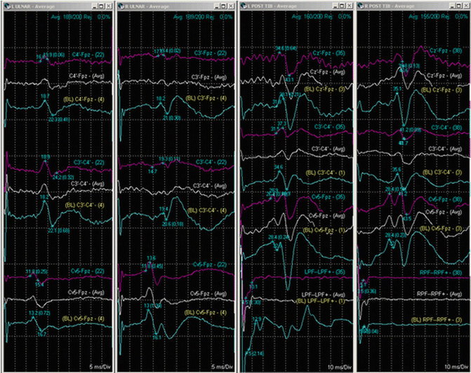
A 67-year-old man with coronary artery disease, diabetes, and hypothyroidism presented with diplopia and was diagnosed with a right-sided sphenoid wing meningioma . He was scheduled for a frontal temporal craniotomy after undergoing preoperative embolization of the tumor a day prior to surgery. Preoperative examination revealed a normal neurological evaluation except for mild diplopia with lateral gaze. The patient was anesthetized with induction boluses of lidocaine, propofol, rocuronium, and fentanyl. The patient was intubated and maintenance of anesthesia was provided by intravenous infusions of propofol, lidocaine, and remifentanil. Additional monitoring included left radial arterial line and a urinary catheter with temperature probe. IOM was established with EMG recordings of cranial nerves III, IV, VI, and VII along with four-limb SSEP. The patient was placed in supine position with slight rotation of the torso. His head was secured with Mayfield pins. A warm-air temperature regulation blanket was placed on the patient. After the surgical area was prepped and draped, baseline EMG and SSEP recordings were normal. The craniotomy proceeded uneventfully with nominal blood loss. As the tumor was being resected, a marked decline in amplitude of the right upper extremity SSEP recordings was noted. Cranial nerve recordings were unchanged from baseline (Fig. 27.2).


Fig. 27.2
SSEP recording from the four extremities with changes in upper right extremity that was related to positioning
Differential Diagnosis of SSEP Findings
The decrease in amplitude of the right upper extremity SSEP recordings prompted an investigation of both a physiological and nonphysiological etiology to the change from baseline. Technical issues related to the monitoring were less likely given that the recordings from the other extremities were unchanged and after verifying the integrity of the electrodes placed in the right upper extremity. The maintenance anesthesia regimen had not been changed nor had the patient experienced significant hemodynamic or temperature variations. Due in part to the preoperative embolization of the tumor, minimal blood loss has occurred at this point, which made it unlikely that anemia was contributing to the changes in amplitude. Furthermore, since the findings were isolated to a single extremity, this suggested that the changes in amplitude were not systemic in nature, but rather directly related to the surgical procedure or to the patient’s positioning. Given that the findings were ipsilateral to the operative site, it was determined that the positioning of the right upper extremity was most likely the cause of changes in SSEP.
Case Progression
After being notified that the right upper extremity SSEP recordings had changed, the surgeon was able to stop operating while the IOM findings were verified and investigated. Examination of the patient’s right arm revealed that it had moved when the Mayo stand had been repositioned during the operation. Once the placement of the Mayo stand was corrected, the SSEP recordings returned to baseline values over the next 20 min. Upon conclusion of the operation, the patient was extubated in the operating room and transported to the intensive care unit. Postoperative examination revealed no sensory or motor deficits in the right upper extremity. Cranial nerve functions were also found to be at preoperative baseline.
Conclusion
In the first case, changes in baseline ABRs were interpreted correctly, reported to the surgeon, and the appropriate action was implemented to resolve the findings. While the sensitivity and specificity of intraoperative ABR changes are often questioned [16, 17], our case outlines an example where utilization of IOM may have prevented unwanted neurological morbidity. This is consistent with other data that suggest that the use of ABR in skull base surgery may improve patient outcomes [18]. In the second case, changes in SSEP recordings were evaluated and lead to an intervention that might have prevented peripheral nerve injury due to an alteration in the patient’s position. While not isolated related to the operative site, positional neuropathies are a common morbidity associated with prolonged surgical times often found with skull base surgeries. IOM during skull base surgery is a valuable tool that benefits both the patient and the practitioner. Correct implementation of various IOM modalities and proper interpretation of their recordings may help with the identification of neuroanatomical structures, the detection of mechanical and thermal injury, and facilitate progression of the surgery.
Stay updated, free articles. Join our Telegram channel

Full access? Get Clinical Tree


