Benefits
Portable and easily accessible. Learners can work through diagnoses at their own pace
Opportunity for self-practice and repetitive practice
Not constrained by patient or instructor availability
Multiple examples of a single diagnosis can be presented quickly and economically
Limitations
Unable to correlate sounds with tactile or visual patient cues. Lack of interaction with physical “patient” (real or simulation)
In the absence of instructor, lack of interaction and feedback regarding auscultatory abnormalities
Audio simulations, which typically consist of CD-ROMs but can include other sound file formats, present recordings of real or simulated patient heart sounds. The benefit of this approach is that the recorded audio sounds are free of contaminating background noise commonly found at the bedside, so it may be easier for the learners to hear the sounds [3]. This is unlike the difficulties encountered when listening to a patient’s heart in a noisy setting, such as the emergency department, where a conventional acoustic stethoscope is unable to filter the additional environmental noise making auscultation more challenging. For novice learners, the absence of extra visual and tactile stimuli may simplify the task at hand and improve their auscultatory accuracy. Supporting this argument, Vukanovic-Criley et al. noted that learners and clinicians at all levels of experience would close or avert their eyes when auscultating a virtual patient that processed a combination of sights and sounds. This was done instinctively, despite the fact that doing so would mean the clinician would actively choose to ignore visual reference that could have assisted them the timing of sounds and murmurs [2].
The efficacy of the audio simulations has been demonstrated in at least two studies [12, 13]. Horiszny demonstrated that repetitive auscultation of heart sounds and murmurs with interactive discussion about pathophysiology with an instructor improved the cardiac auscultatory proficiency of family medicine residents [13]. Auscultatory diagnostic accuracy improved from 36 to 62% after three 45-min sessions, as tested using the simulated sounds. A similar study confirmed the utility of the audio recordings for medical students after a 2½-h session with an instructor [12]. These studies demonstrate the effectiveness of repetitious listening to heart sounds when enhanced by the presence of an instructor providing feedback and guiding group discussion.
Another study powerfully demonstrated the importance of repetitive listening to heart sounds and murmurs [14]. Third-year medical students either listened to 500 repetitions of 6 simulated heart sounds and murmurs, recorded on a 1-h CD, or were not exposed to any recorded sounds. Those students who engaged in repetitive practice had significantly higher diagnostic accuracy on both simulated and real patient heart sounds.
These studies support the efficacy of pure auscultatory simulations in improving diagnostic accuracy, at least as tested on auscultatory simulations. These types of simulators are well suited to novice learners and require only a few hours of repetitive practice to demonstrate significant learning gains.
Multimedia Simulations (Table 18.2)
Table 18.2
Multimedia simulators
Benefits |
Able to present interactive cases with additional supportive history, laboratory tests, and explanations of pathophysiology |
Incorporation of visual stimuli, such as ECG tracings, pulse waveforms, videos of carotid pulses, JVP, and precordium, to help correctly locate and identify heart sounds and murmurs |
Opportunity for self-practice and repetitive practice |
Not constrained by patient or instructor availability |
Limitations |
In the absence of instructor, limited feedback from computer (absence of feedback with non-CAI multimedia formats) |
Lack of interaction with physical “patient” (real or simulation) |
More expensive and less accessible than exclusively audio simulations |
CD-ROM
It has been hypothesized that the lack of information regarding location, intensity, and radiation of the murmur may limit the utility of the exclusively audio simulations [14]. Critics of these simulators suggest that listening to heart sounds and murmurs in isolation without visual and tactile stimuli is not reflective of auscultation at the bedside [3]. Multimedia cardiac simulation typically involves audio recordings of heart sounds with visual stimuli in the form of graphics, ECG tracings, phonocardiograms, and video recordings of the jugular venous pulsation (JVP), carotid arteries, and precordium. Additionally, some of these simulations have supplementary history, imaging (e.g., CXR, echocardiogram), and teaching points. The primary characteristic of multimedia cardiac simulations is the ability to identify systole and diastole through the visual modality, allowing the user to properly time auscultatory findings within the cardiac cycle.
Multimedia CD-ROMs have been shown to improve cardiac auscultation knowledge and skill [3, 6, 15, 16]. The study designs include a single-group pre-post study [16], cohort studies comparing multimedia to classroom teaching [15, 16], and multimedia plus traditional clerkship to clerkship alone [6, 15]. For example, Stern et al. [6] found that students who were exposed to CD-ROM cases demonstrated improved diagnostic accuracy when tested on simulated sounds compared to students who only experienced the traditional clerkship instruction. Furthermore, the subset of students who had the full intervention (reviewing in-depth CD-ROM cases and 20 mini-cases) had a preservation of auscultatory skill when retested 9 months later.
In the study by Stern et al., and in another study by Finley et al. [6, 15], students used the multimedia simulations either on their own or in the presence of an instructor. Both studies demonstrated similar diagnostic accuracy on simulated heart sounds between both instructor present and instructor absent groups. These studies suggest that students can use these simulations independently and demonstrate significant gains in skills. However, from the students’ perspective, they felt that a combination of both classroom and computer learning would be preferable to either modality alone [15].
Computer-Assisted Instruction
For our purposes, computer-assisted instruction (CAI) is defined as a multimedia simulation program that uses computer programs or Web-based learning as the central means of information delivery but is also interactive. Within cardiology, considerable interest in CAI has led to the development of CAI modalities for teaching cardiac physiology and cardiac physical examination skills. Conceptually complex, visually intense, and detail-oriented tasks such as understanding the pathophysiology behind a murmur are well suited to CAI [15]. An interactive computer screen can show animations of anatomy, ECG tracings, pressure-volume curves, and echo images, in addition to supplementary text or audio instruction to help the learner acquire the necessary background knowledge. This can then be coupled with heart sound recordings and videos of physical examination to serve as a comprehensive presentation of a particular cardiac condition. Similar to the multimedia CD-ROMs, CAI allows the student to engage in self-directed learning in a nonthreatening environment [17]. Both multimedia modalities generally do not require immediate direct supervision from the instructor, thus reducing the geographical and time constraints on both students and instructors [17].
Perhaps the best studied example of a cardiology-specific CAI is the UMedic system (University of Miami medical education development system for instruction using computers) [18, 19]. Developed to be used in conjunction with Harvey® The Cardiopulmonary Patient Simulator, UMedic is a multimedia program that presents an extensive cardiovascular curriculum, incorporating cardiac auscultation and cardiovascular imaging in its presentation [20] (Fig. 18.1). It has been shown to improve diagnostic accuracy on simulated cardiac findings, for clerkship students, compared to a traditional clerkship rotation [19].
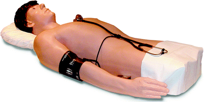
Fig. 18.1
Harvey® The Cardiopulmonary patient simulator (Photo courtesy of Laerdal)
Sverdrup et al. [21] addressed whether traditional bedside teaching versus training with CAI would lead to differences in diagnostic accuracy with real patients. Two groups of third-year medical students received a 2-h instructional session focused on cardiac examination and physiology. One group received an additional 2-h traditional bedside teaching session, while the other group went through a series of cases with the multimedia simulation. Both groups had equal diagnostic accuracy when tested with real patients.
Virtual Patient Encounters
Virtual patient encounters (VPE) are defined as “a specific type of computer program that simulates real-life clinical scenarios; allowing learners to obtain a history, conduct/view a physical exam, assess diagnostic tests and make diagnostic and therapeutic decisions” [21, 22].
These interactive multimedia teaching programs use real or standardized patients filmed at the bedside with supplemental animations, demonstrations of anatomy, ECG tracings, echocardiogram images, and text or audio instruction. In addition to the ancillary information, actual recorded heart sounds and actual video recordings of a live patient are presented. The VPEs permit the learner to move the virtual stethoscope over the virtual patient’s precordium while observing pulses, respiration, and/or postural maneuvers [7].
Virtual patient encounters can improve the cardiac examination competency of medical students [7]. In one study, 24 medical students received eight 90-min sessions with a VPE in addition to their baseline core curriculum and were compared to 52 students receiving no additional instruction. The VPE group improved their diagnostic accuracy when tested on the VPE compared to the control group. Moreover, a subset of students in the intervention group was tested 14 months later and had a sustained increase in accuracy compared to the control group. VPEs have also been used to assess the knowledge and auscultation skills of medical students, residents, fellows, and clinicians [2].
Despite the appeal of multimedia simulations, whether CD-ROM, CAI, or VPE, there still exists a need for more well-designed studies examining how best to use these simulation modalities. The majority of studies employ nonrandomized designs, add multimedia simulators to instruction received by both groups as opposed to comparing two interventions that require equal time, and test learners using the multimedia simulator as opposed to real patients. Without stronger research designs, and particularly without demonstrating the translation of skills from simulation to real patients, the benefits of multimedia simulations remain under-explored.
Standardized Patients (Table 18.3)
Table 18.3
Standardized patients
Benefits |
Real person with which to interact |
Ability to learn the techniques and cardiac findings of the normal cardiac physical examination |
Patient and/or instructor present to provide feedback on performance |
Potential for “hybrid” simulations, combining the normal physical examination of the patient with any of the simulation modalities presenting the cardiac abnormalities |
Limitations |
Typical standardized patient does not have cardiac abnormalities, thus unable to present abnormal physical findings |
Inefficient, as only a few students can examine the patient at any one time |
Not conducive to repetitive practice or available for independent practice |
Cost and time intensive to recruit and train appropriate standardized patients |
A standardized patient (SP) (see Chap. 13) is an actor or patient who has received training to present his or her history in a standardized, reliable manner and who sometimes mimics physical signs [10]. The use of SPs for teaching the basics of cardiac physical examination is widespread in North American undergraduate medical education, as is their use in assessment [11]. SPs may be helpful for teaching normal physical exam findings and instructing learners in physical exam technique. However, the SP’s normal physical exam poses a problem when assessing learners’ ability to recognize abnormal clinical signs and to apply and integrate knowledge [23]. There is a low correlation between clinicians’ physical examination technique and their ability to diagnose cardiac abnormalities [24].
Lecat’s Ventriloscope, manufactured by Limbs & Things, is a modified stethoscope that allows prerecorded sounds (activated wirelessly) to be integrated with a standardized patient [23]. It is designed to overcome some of the limitations of the SP with normal physical findings. The Ventriloscope allows for projection of abnormal auscultatory signs onto a healthy person, requiring students to recognize and interpret such signs within the wider context of a clinical encounter [25]. The learner also benefits by completing the cardiac examination on a live person. Additionally, the same SP can have a number of cardiac conditions that can be interchanged with ease (Fig. 18.2).
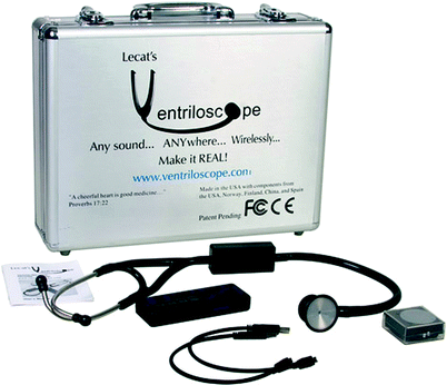
Limitations of early Ventriloscope models included lack of synchronization of cardiac auscultatory findings with the SP’s pulse and lack of respiratory variation of the heart sounds. Technology is being developed to track the pulse (by having the SP wear a heart rate monitor) and trigger the recorded sounds simultaneously. A foreseeable limitation to the Ventriloscope is that it simulates auscultatory abnormalities, but cannot simulate associated physical examination findings, such as JVP or pulse abnormalities. The Ventriloscope is a relatively new technology, and comparative effectiveness studies are not presently available.
For teaching cardiovascular physical examination, perhaps the ideal use of the SP is to combine the teaching of the normal cardiac examination with recognition of auscultatory abnormalities using any one of the simulation modalities. Although such “hybrid” or integrated simulations are being used in other domains, especially invasive procedural skills training [20, 23, 26–29], and despite the prevalent use of SPs in the medical education system, there is little research examining how best to use them for cardiac physical examination teaching.
Cardiopulmonary Simulators (CPS) (Table 18.4)
Table 18.4
Cardiopulmonary simulators
Benefits |
Simulate physical patient, with palpable pulses, JVP waveforms, precordial movements, and simulated heart sounds. Facilitates comprehensive physical examination as would be performed on a real patient |
Allows for repetitive practice and can provide opportunity for self-practice |
Typically, instructor present to provide feedback on performance |
Multiple learners can listen to and examine the simulator at the same time, without the issues of real patient fatigue |
Limitations |
More expensive and less accessible than other simulator modalities. A single institution may have only one cardiopulmonary simulator, thus limiting learner access to the simulator |
Typically only have one or two examples of each diagnosis |
Although self-study modes are available, typical presence of instructor is faculty intensive |
Cardiopulmonary simulators (CPS) are mannequin-based simulators that have palpable pulses, JVP waveforms, precordial movements, and simulated heart sounds. Conceptually, CPS have great potential as tools to enhance the education of learners’ cardiac examination skills. By more closely mimicking a real patient, CPS allow a full cardiac examination, integrating all sensory information. Cardiopulmonary simulators can replicate abnormal pathology and can be reviewed at the instructor’s and learner’s convenience, thus alleviating the constraints of patient and pathology availability. Unlike real patients who may tire from being examined by multiple trainees, multiple learners can listen to and examine the simulator at the same time, thus increasing the efficiency of teaching.
Almost all of the research into the effectiveness of CPS as a teaching modality has been undertaken using Harvey®. Across multiple studies, it has been demonstrated that instruction with Harvey®, either in isolation or in conjunction with the UMedic multimedia system, can improve novice and resident learners’ diagnostic accuracy on Harvey® and on real patients [20, 23, 26–29]. Typically, these are single-group studies, but one was a cohort design comparing a fourth-year medical student cardiology elective focused on Harvey® examination in addition to real patients to a traditional elective focused on real patients. There was a small but statistically significant superiority in diagnostic accuracy with real patients for the Harvey®-trained students [28].
There has only been one study that compared CPS to another simulation modality for teaching cardiac physical examination skills [30]. This study suggested that instruction with Harvey® was no more effective than instruction using CD-ROMs [30], but limitations of the study design including no pretest to establish equivalence of the two groups at baseline and difficulty establishing equivalence of the posttests done with real patients limit interpretation of the results. In one study using only CPS, medical students demonstrated a transfer of skills from CPS to real patients for a cardiac murmur presented on the CPS but a lack of transfer from simulated murmurs that were different from the real patient’s diagnosis [31]. Thus, the comparative benefit of CPS over other simulation modalities remains largely unexplored.
Related posts:


Stay updated, free articles. Join our Telegram channel

Full access? Get Clinical Tree


