Fig. 21.1
Simulated head with torso (Used with permission from the KaVo Group)
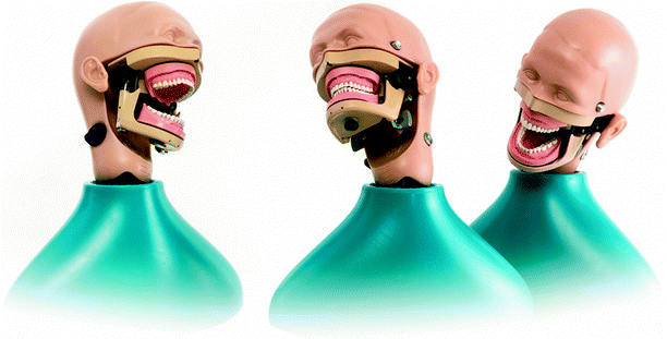
Fig. 21.2
Simulated head and torso with masticatory system (Used with permission from the KaVo Group)
Standardized Patients
Standardized patients [17] (see Chap. 13) have been used in medical education since the early 1960’s and were first reported in dental education in 1990 [18]. Standardized patients are very helpful in developing appropriate communication and interpersonal skills for dental students, eliciting a thorough medical and dental history, completing a head and neck exam, and completing accurate and appropriate record keeping. Unfortunately, standardized patients are not available at many health science centers and if available, may be cost prohibitive for many dental schools.
Web-Based and Computer-Based Simulation
Case-Based Simulations
Computers can be extremely helpful in simulating patients or presenting case scenarios [19–22]. In case-based computer programs, students are presented a narrative that represents a patient encounter. The students can interact with the program that provides responses to a variety of interventions including answers to a series of questions relating to medical history, diagnostic test results, orders, possible diagnoses, and the development of appropriate treatment plans. These programs are becoming more and more interactive, allowing for more pertinent feedback to the student.
Simulation-based Assessment
Since the early 1990s, computer simulations have been considered for dental licensure examinations or other evaluations and testing applications; however, when used for these purposes, reliability and validity are of utmost concern [23]. The goal when using simulation for education is to achieve performance improvement, whereas simulation for dental licensure examinations is used to ensure that the provider has reached competency. This requires that simulation for these high-stakes assessments must meet a higher standard in reliability and criterion validity. The advantages, however, of using simulation for dental examinations, assuming the level of reliability and validity is sufficient, are compelling. Simulation may have a greater potential validity, is safer in comparison to using live patients, and may be less expensive and easier. Some schools use simulation to assess students during their education, but advanced simulation is not currently used in any clinical examination for state licensure. As simulations in dentistry become more and more advanced, their use in dental licensure examinations will receive more consideration.
Computer Imaging
Recent advances in computer imaging have allowed for the production of very accurate 3D dental models of the oral apparatus by destructive scanning, laser scanning, or direct imaging of teeth [24–27]. Also imaging systems have been used to capture 3D images of mandibular motion [25]. The technology of computer imaging is important in the educational component of dentistry since it can dictate the relation to reality. Computer imaging is instrumental in simulations using virtual reality and/or haptics. Accurate and detailed imaging is critical because dental procedures require measurements in the tenths of millimeters; hence, virtual images must have very high resolution to order to evaluate such precise detail.
Virtual Reality-Based Technology (VRBT)
DentSim System
Mannequin-based simulation has become the standard of preclinical teaching in dental schools as described at the beginning of this chapter. Over a decade ago, a computerized dental simulator was developed by DenX Ltd. (presently owned by Image Navigation Ltd., Fig. 21.3). This new virtual reality simulator allows students to receive immediate, three-dimensional, audio, and written feedback of their work on artificial teeth (such as cavity, crown, and endodontic access preparations) and to review their work on video. This computerized simulator can be installed on an existing traditional mannequin, with the addition of a computer, camera, a special handpiece (dental drill), and a reference body that functions as the tracking device [2].
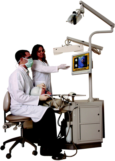

Fig. 21.3
Virtual reality-based simulator (DentSimTM, Image Navigation Ltd. Used with permission)
The DentSim tracking system technology is based on the principles of GPS (global positioning system) technology. The optical system uses a camera to track a set of light-emitting diodes (LED), located on the dental handpiece and on the simulator’s jaw. The LEDs send infrared signals that are picked up by the camera which is located above the workstation. The handpiece LEDs provide data indicating the motions of the user, while the LEDs attached to the jaw provide data regarding the location of the teeth. Once the camera has picked up the signals, the exact location of the tip of the bur (dental drill bit) in relation to the tooth may be calculated. This information is transferred to the computer and displayed through virtual simulation and as a color-coded, three-dimensional, tooth image.
Using the virtual reality simulator, the student’s work is recorded and compared to an ideal preparation that is predesigned or selected by the course director from the software database. The students can view an accurate image of the ideal preparation, the student’s preparation, and the exact measurements of each preparation in every dimension.
Several studies have been conducted to test the validity of the DentSim system. This is the only technology of its kind that has been evaluated in the scientific literature. One study demonstrated that when trained with the virtual reality simulator, starting at a minimum of 6 h dental, students learn faster than their peers in the traditional preclinical course, arrive at the same level of performance as expected of students trained in a traditional manner, accomplish more practice procedures per hour, and request more evaluations per procedure or per hour than in the traditional preclinical laboratories [28]. Another study suggested that virtual reality technology has the potential to provide an efficient and more self-directed approach for learning clinical psychomotor skills. That study showed that students using only traditional instruction received five times more instructional time from faculty than did students who used the virtual reality simulation system. There were no statistical differences in the quality of the preparations [29]. Several studies have shown an improvement in course performance through higher examination and course scores [30–32] as well as a decrease in overall course failure rate and elimination of student remediation by more than 50% [31, 32]. In addition, studies have shown the advantage of dental students using virtual reality simulators to learn psychomotor skills, as well as examined faculty members’ evaluation and expectations of employing this relatively new technology [33]. Results for other studies support the use of VRS in a preclinical dental curriculum. This virtual reality-based technology is currently being used in several dental schools around the world (Fig. 21.4). This system has many capabilities which allow specific adaptation and curricular integration based on the individual school’s requirements.
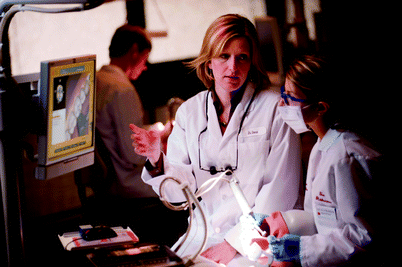

Fig. 21.4
Advanced Simulation Clinic, University of Minnesota, School of Dentistry
EPED System
EPED stands for efficiency, professionalism, education, and dependability. EPED’s main product is the Computerized Dental Simulator (CDS-100), which combines a simulation system, an evaluation system, and 3D virtual reality technology [34]. CDS-100 system is based on exactly the same approach as the DentSim and comprised of a dental handpiece for drilling a cavity in a plastic tooth; a three-dimensional sensor with six degrees of freedom attached to the dental handpiece, which provides the system with the position and orientation of the handpiece; and a data processing and display unit for displaying the procedure [34, 35] (Fig. 21.5). There is no scientific evidence of its use at this time; however, it is expected to have similar educational benefits as the DentSim system.
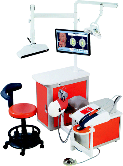

Fig. 21.5
CDS 100 virtual reality simulator by EPED (Imaged used with permission)
Second Life
Dental education has been assessing the value of distance education and the steps required to deliver technology-based learning while providing high-quality patient care teaching methods for dental and dental hygiene students. Second Life (SL) is a three-dimensional technology that provides simulation-based virtual settings. Activities in SL provide a way to combine new simulation technologies with role-plays to enhance instruction in diagnosis and treatment planning. Case studies and role-plays have been used as effective evaluation mechanisms to foster decision-making and problem-solving strategies in the delivery of patient care. It is a newly emerging technology and its benefits are yet to be reported in the scientific literature [36].
Technology with Haptic Focus
The growth of computer hardware and software has led to the development of virtual worlds that support the field of advanced simulation. Virtual reality (VR) creates virtual worlds using mathematical models and computer programs, allowing users to move in the created virtual world in a way similar to real life. Adding haptic technology to the VR world provides users with the ability to interact with virtual objects within the virtual environment via feel and touch. A dental training system using a haptic interface requires haptic rendering to generate force feedback and drilling simulation for the material cutting behavior from the virtual tooth model.
Haptic dental trainers enable students to learn and practice in a safe and almost real environment. Whereas traditional simulation laboratories restrict skill development to practicing on unrealistic plastic teeth, a virtual simulation lab offers the opportunity to let students learn in a realistic, pathology-oriented basis. A large variety of realistic clinical problems can be simulated in the virtual environment to assure the acquisition of the essential competencies to manage a multitude of pathologies. Problem solving, treatment planning, as well as performing treatment can also be learned in this environment before the students encounter actual patients in the dental clinics. Introduction of a virtual clinical lab decreases the gap between the traditional preclinical curriculum and the clinics (Fig. 21.6) by adding more realistic scenarios in the preclinical training environment. The realistic scenarios embed integration of the theory in the preclinical phase and stimulate further theoretical inquiry as a result of virtual clinical experiences.
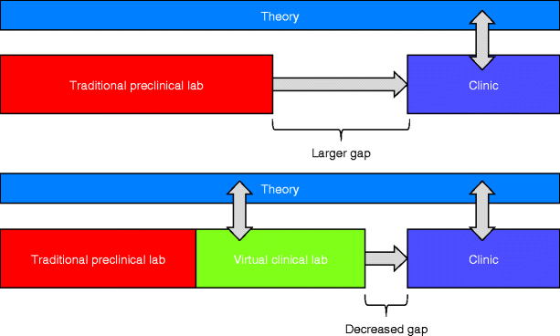

Fig. 21.6
The virtual clinical lab integrates theory and decreased the gap to the clinic
In the virtual clinical lab, students can encounter and provide treatment to a variety of simulated patients with dental pathology. Although the use of actual extracted teeth in the preclinical simulation lab may add some reality and pathology, it is impossible to offer students similar cases and scenarios without virtual capabilities. Besides, it becomes clear that current restrictions on the use of extracted teeth as well as their limited availability in some regions make their use more difficult. Because of this shortage of suitable extracted teeth, it is hard for students to train and assess all skills taught in the preclinics.
Another advantage of the virtual clinical lab is the ability to combine the standardization of exercises, as the students would experience in the present preclinical environment, with the realistic scenarios as encountered in the clinic. This ability provides an excellent environment for competency development. Other VR-related advantages include the ability to assess complete processes instead of the end result, while allowing students the opportunity to practice exercises without limits and without the need to use exhaustible materials like drills and plastic teeth (Fig. 21.7).
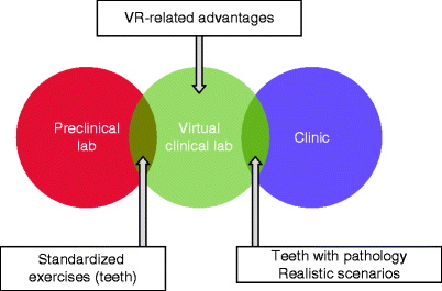

Fig. 21.7
The virtual clinical lab features advantages of both the preclinical lab and the clinic
The virtual lab also has some limitations. Not all procedures are equally suitable for training in a virtual clinical lab. For instance, applying filling material, matrix band, and wedge procedures might be hard to train virtually. Also feeling and handling of teeth in virtual reality is not as rich and versatile as it is in the real world. The virtual clinical lab should be considered an adjunct to the learning potential of the existing preclinical lab, not a replacement. Combining them provides the opportunity to use the best of both worlds in preparing for the clinics.
Recently, VR simulators using haptic feedback have been introduced into the dental curriculum as training devices for manual dexterity acquisition in tooth probing and preparation tasks. Various systems have been developed of which a few are on the market or entering the market [37–40].
PerioSim is an example of a haptic technology system that uses a periodontal probe [37]. It was developed at the University of Illinois at Chicago through joint efforts of the College of Dentistry and the College of Engineering. The system consists of a high-end computer workstation, a haptic device, and a stereoscopic computer monitor with stereo glasses. It was developed for training and evaluation of performance by students in periodontal probing and detecting the feel of a caries-active white spot lesion.
A realistic 3D human mouth is shown in real time, and the user can adjust the model position, viewpoint, and transparency level. The haptic device allows the student to feel the sensations in the virtual mouth. The instrument pressure (in grams of force being applied to the gingival area) can be viewed and recorded. A control panel is available for fine control of a variety of parameters such as instrument and model selection, degree of model transparency, navigation, haptic fidelity of tissues, and tremor modulation.
The system allows instructors to create short scenarios of periodontal procedures, which can be saved and replayed at any time. The 3D component permits students to replay the procedure from any angle. This allows the user a unique advantage to observe different views of the placement of the instrument and gingival relationships during a given procedure. It appears that the PerioSim is considered as highly realistic with proper feedback for teeth and instrument [41].
VOXEL-MAN Dental [38] was developed by the VOXEL-MAN group within the University Medical Center Hamburg-Eppendorf. The VOXEL-MAN Dental (Figs. 21.8 and 21.9) is a compact unit that comes in a variety of configurations including a tabletop system, a stand-alone system, or attached to a conventional dental training unit. With this device teeth and instruments are computer generated via high-resolution modeling displayed on a 3D screen. The high-resolution tooth models were derived from real teeth by microtomography. The dental handpiece can be moved in three dimensions and is represented by a force feedback device. This interface provides a very lifelike and convincing sense of touch. Impressively the differences in feel between enamel, dentin, pulp, and carious tissue have been faithfully reproduced.
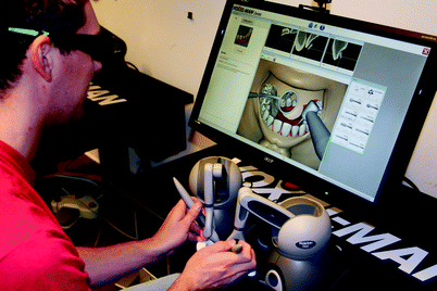
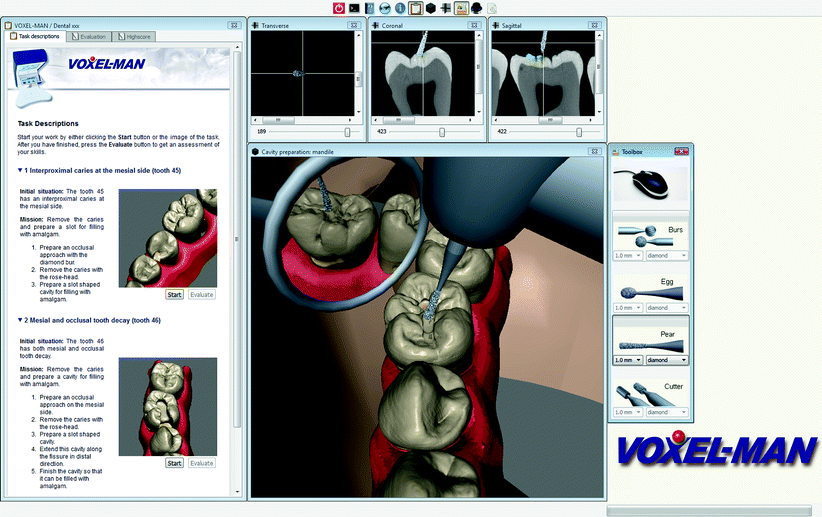

Fig. 21.8
The VOXEL-MAN tabletop system

Fig. 21.9


Screen shot of the VOXEL-MAN system. (Used with permission from Springer)
Stay updated, free articles. Join our Telegram channel

Full access? Get Clinical Tree


