Fig. 19.1
Small vessel anastomosis: (a) An anastomotic block can be constructed using a wood block on which are mounted ¼ in. angled irrigation connectors. A synthetic graft can be positioned in place using the connectors (Reprinted from Fann et al. [3], with permission from Elsevier). (b) Mounted in the portable chest model is a synthetic target vessel; to simulate vein graft for anastomosis, another synthetic vessel is used. (c) For the tissue-based or “wet-lab” component, porcine hearts are prepared and positioned so as to expose the left anterior descending artery in a container
Beating Heart Surgery
The technical challenge of off-pump coronary artery bypass grafting is to expeditiously perform accurate coronary anastomoses on constantly moving target vessels, typically 1–2 mm in diameter. Understanding the stabilization devices is critical as is the various methods of optimizing exposure of the target vessel [2]. Commercially available synthetic beating heart model includes a simulator from the Chamberlain Group, which includes a compressor and controller (Fig. 19.2a), and one developed by EBM, which is motor driven (EBM, Tokyo). Tissue-based models using explanted porcine hearts include the Ramphal simulator, which is a high-fidelity, computer-controlled simulator that can be employed in many aspects of training (Fig. 19.2b). This educational approach permits the surgeon to become familiar and achieve some degree of proficiency before attempting this technique in the clinical setting.
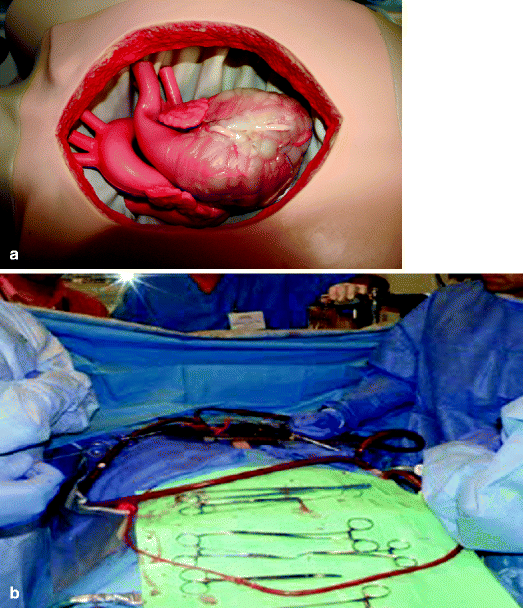
Fig. 19.2
Beating heart simulator: (a) The beating heart model is constructed of silicone and connected to a controller and external compressor. Partially embedded in the myocardium are 2-mm target coronary arteries. The heart is placed in a plastic torso simulating the pericardial well. (b) The Ramphal cardiac surgery simulator is a high-fidelity computer-controlled tissue-based simulator that allows the trainee to perform tasks such as beating heart and arrested heart surgery. Additionally, it provides cardiopulmonary bypass simulation and crisis management
Aortic Cannulation
One part-task simulator for aortic cannulation is the HeartCase model with the synthetic thoracic aortic attachment. Using a syringe or a pressure bag, normal saline can be instilled into the synthetic aorta for pressurization. A tissue-based model used at the Boot Camp is a porcine heart in which the coronary sinus, coronary arteries, and aortic arch vessels are oversewn (Fig. 19.3a) [5, 28]. The ascending aorta with the arch and a portion of the descending aorta is pressurized with a bag of saline. Another model utilizes the porcine descending thoracic aorta, which is prepared by oversewing the intercostal vessels, securing it in a plastic container, and pressurizing it using saline (Fig. 19.3b) [4]. The tissue-based models are realistic, permitting multiple cannulations. Also, they can serve as thoracic aortic surgery anastomosis simulators [4].
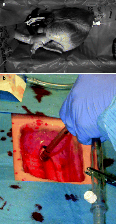
Fig. 19.3
Aortic cannulation: (a) For the perfused non-beating porcine heart placed in a container, the arch vessels are oversewn, and a portion of the descending aorta in continuity with the ascending aorta provides a long segment to practice multiple aortic and cardioplegia cannulations. (b) A porcine descending thoracic aorta is secured in a plastic thoracic model. The pressurized aorta allows placement of purse-string sutures and multiple aortic cannulations (Reprinted from Fann et al. [4], with permission from Elsevier)
Atrial Cannulation
Right atrial or bicaval cannulation for cardiopulmonary bypass is simulated using the porcine heart model placed in a container. Understanding the anatomy, suture placement, and cannulation are the primary objectives. Ideally, atrial cannulation is performed as part of other procedures, such as aortic cannulation. In order to simulate atrial cannulation in a more realistic, beating heart setting, the Ramphal simulator can be used.
Cardiopulmonary Bypass
Used in the training of perfusionists and surgeons, cardiopulmonary bypass simulators are intended to be highly interactive [4, 5]. The simulators provide multiple physiologic conditions and permit the trainee to manage the steps preceding and during cardiopulmonary bypass, including intraoperative crisis management. The Ramphal simulator is thus well suited for cardiopulmonary bypass simulation. Other “high-tech, high-fidelity” cardiopulmonary bypass simulators, such as the Orpheus perfusion simulator (ULCO Medical), are commercially available with assessment modules, recognizing that such simulators are expensive (Fig. 19.4). A commonly employed method to learn about cardiopulmonary bypass circuit is to have access to a perfusion pump and arrange a tutorial with the perfusionist.
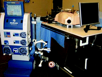
Fig. 19.4
Cardiopulmonary bypass: The Silastic heart model is placed in a plastic thorax and attached to the Orpheus cardiopulmonary bypass simulator. This simulation exercise allows the trainee to understand the cardiopulmonary bypass circuit and to participate in emergency and crisis management (Reprinted from Hicks et al. [5], with permission from Elsevier)
Aortic Valve Replacement
Synthetic models are adequate in teaching important components such as surgical anatomy and approach to placing annular sutures [4]. The aortic root model is available from the Chamberlain Group and can be attached to the HeartCase simulator (Fig. 19.5a). The objectives are to train proper needle angle for suture placement, effective knot tying in a deep confined space, placing sutures in the valve sewing cuff, and seating the valve prosthesis. For tissue-based simulation, porcine hearts are placed in a container and situated so as to present the ascending aorta and aortic root (Fig. 19.5b) [4]. The aortotomy is made followed by excision of the leaflets and the muscle bar under the right coronary cusp. Interrupted sutures are placed followed by the placement and seating of the prosthesis. This model lends itself to standardized training, as it is currently used in many centers [4].
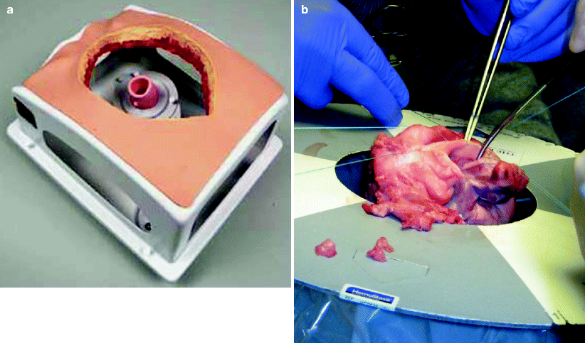
Fig. 19.5
Aortic valve replacement: (a) Mounted in a portable HeartCase chest model is a silicone-based aortic valve model which requires the trainee to understand proper needle angles, working in a deep, confined space, and seating the prosthesis. (b) For the tissue-based aortic valve replacement model, a porcine heart is placed in a container and situated so as to present the ascending aorta and aortic root. The aortotomy is made followed by excision of the leaflets and implantation of the aortic valve (Reprinted from Fann et al. [4], with permission from Elsevier)
Mitral Valve Repair
The synthetic mitral valve attachment is a silicone-based cylinder placed in the portable HeartCase model (Fig. 19.6a) [4, 6]. The synthetic model provides a method to learn basic components of mitral valve surgery, such as exposure techniques and needle angles, but it is limited in its fidelity [4]. To more accurately simulate the mitral leaflets and annulus requires the use of a porcine heart model, which is placed in the container and situated so as to present the left atrium and mitral annular plane. The left atrium is opened and the mitral valve and annulus exposed in an “anatomically correct” configuration (Fig. 19.6b) [4, 6]. Interrupted annular sutures are placed and annuloplasty ring situated and secured. The porcine model is realistic but can pose some challenges with anterior-posterior orientation in the setup [4].
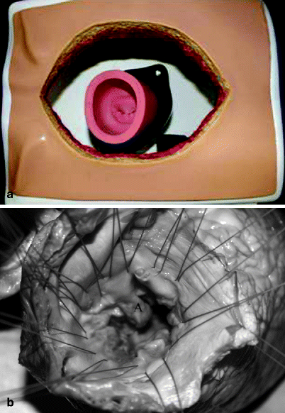
Fig. 19.6
Mitral valve repair: (a) The synthetic mitral valve model is placed in portable chest model. (b) For the tissue-based simulator, porcine hearts are placed in the container and situated so as to present the mitral valve. The left atrium is retracted so as to expose the mitral valve and annuloplasty performed. A anterior leaflet (Reprinted from Joyce et al. [6], with permission from Elsevier)
Aortic Root Replacement
For the tissue-based aortic root replacement simulator, explanted porcine hearts are placed in the container and situated so as to present ascending aorta and aortic root [4]. The porcine aorta and root are resected after creation of the coronary ostial buttons. A composite valve graft (or just a Dacron graft) or an expired aortic homograft (CryoLife, Inc. Kennesaw, GA), if available, is prepared and anastomosed as a root replacement using polypropylene sutures for coronary button reimplantation [4]. This realistic, tissue-based model has been helpful in familiarizing the trainee with the complexity of this procedure [4].
Thoracic Surgery Simulators
Hilar Dissection and Pulmonary Resection
A porcine heart-lung block placed within the chest cavity of a mannequin simulates the necessary maneuvers of hilar dissection and pulmonary resection through a thoracotomy incision (Fig. 19.7) [4, 11]. Either the right or left lung can be used. This tissue-based simulator replicates the confined space in which pulmonary resections are performed and provides a method to practice hilar dissection and resection skills. The objectives are to identify anatomic landmarks, dissect and encircle the hilar vessels and bronchus, and ligate and divide vascular structures using sutures and staplers. This model is moderately realistic recognizing variability of porcine anatomy relative to human anatomy and the fragility of the vascular structures [4].
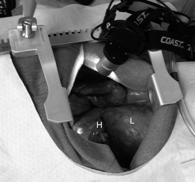
Fig. 19.7
Hilar dissection: a porcine heart-lung block placed within the chest cavity of a mannequin simulates the necessary maneuvers of hilar dissection through a thoracotomy incision. H hilum, L lung (Reprinted from Fann et al. [4], with permission from Elsevier)
Esophageal Anastomosis
Placed within a thoracic mannequin, a porcine heart-lung-esophagus block simulates the thoracotomy providing access to the posterior mediastinum [4]. The esophagus, positioned and secured in the posterior cavity, is isolated and transected. The two free ends are re-approximated in either one or two layers. This model permits alignment and approximation of the esophageal ends, proper placement of sutures within the esophageal wall, and securing the sutures following placement [4]. By including the stomach in the tissue block, esophagogastric anastomosis can be simulated. Additionally, providing and resolving tension on the anastomosis can be introduced, and creating longitudinal incision (with longer mucosal than muscular incision) would simulate esophageal rupture requiring the trainee to perform appropriate repair [4].
Rigid Bronchoscopy
Using conventional bronchoscopic equipment, the TruCorp AirSim simulator (Belfast, N. Ireland) is a model of the oral pharynx, larynx, and the tracheobronchial tree out to the segmental anatomy (Fig. 19.8) [4]. The model also allows simulation of awake bronchoscopy, bronchial stent placement, and removal of foreign body [4]. Since the image of the bronchoscope can be projected and the position of the light on the bronchoscope can be visualized through the wall of the model, assessment of resident performance in navigation is possible. Another bronchoscopic simulator is the CAE Accutouch EndoscopyVR Surgical Simulator (CAE, Montreal, Quebec), which is a highly sophisticated virtual reality simulator.
Fig. 19.8
Bronchoscopy: using conventional bronchoscopic equipment, the TruCorp AirSim model simulates the tracheobronchial tree
Video-Assisted Thoracoscopic Surgery (VATS) Lobectomy
A left porcine heart-lung block placed within the chest cavity of a mannequin is accessed via working ports to allow for video-assisted resection (Fig. 19.9) [4]. This exercise replicates the confined thoracic space in which pulmonary resections are performed and also provide a model to practice hilar dissection and resection skills. This simulator allows for identifying anatomic landmarks, maneuvering the thoracoscope and pulmonary structures, dissecting and encircling hilar vessels and bronchus, and dividing the structures using the endoscopic staplers. Recognizing interspecies differences, this model may be more complex than a case in the clinical setting, but it does provide simulation of many advanced maneuvers [4].
Fig. 19.9
Video-assisted thoracoscopic surgery (VATS) lobectomy: a left porcine heart-lung block placed within the chest cavity of a mannequin is accessed via working ports to allow for video-assisted resection (Reprinted from Fann et al. [4], with permission from Elsevier)
Tracheal Resection
A porcine tracheal-esophageal segment placed within the open neck of a mannequin simulates tracheal resection and anastomosis (Fig. 19.10) [4]. This realistic exercise reproduces the confined space in which tracheal resections are performed and provides a model to practice such resection and anastomosis. Given the shorter period of time required for this exercise, additional procedures, such as tracheostomy and tracheal release maneuvers, can be added [4].
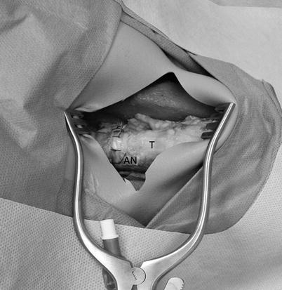
Fig. 19.10
Tracheal resection: a porcine tracheal-esophageal segment placed within the open neck of a mannequin simulates tracheal resection and anastomosis (T trachea, An anastomosis) (Reprinted from Fann et al. [4], with permission from Elsevier).
Sleeve Resection
As an extension of the pulmonary resection simulator, a porcine heart-lung block placed within the chest cavity of a mannequin simulates the necessary maneuvers of sleeve resection via a thoracotomy (Fig. 19.11) [4]. This exercise replicates the confined thoracic space in which sleeve resections are performed. This realistic model permits airway mobilization and understanding the principles of bronchial anastomosis [4].
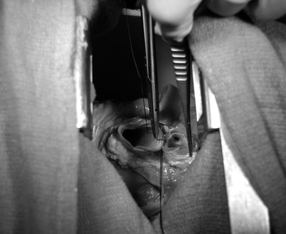


Fig. 19.11
Sleeve resection: a porcine heart-lung block placed within the chest cavity of a mannequin (similar to the hilar dissection model) simulates the necessary maneuvers of sleeve resection (Reprinted from Fann et al. [4], with permission from Elsevier)
Related posts:


Stay updated, free articles. Join our Telegram channel
Full access? Get Clinical Tree
