Respiratory Complications Overview


Respiratory Complications Overview


Robert R. Kirby


Why Do Pulmonary Problems Occur?

Pulmonary complications are the most frequently reported causes of postoperative morbidity and mortality,1,2 in particular, after procedures involving the abdomen and thorax.3,4 The incidence reportedly is as high as 75% in some patient populations.5,6 Yet, as the case summary shows, they may occur in the absence of major surgical procedures when one might least expect them. When possible, the identification of patients at risk—to allow appropriate interventions that limit that risk—is imperative before surgery. However, in this case, no amount of preanesthetic assessment would have indicated that this perfectly normal and healthy young man would develop problems that would convert his anticipated outpatient surgical procedure into a 3-day hospital stay.


▪ RISK FACTORS

Prolonged mechanical ventilation, pneumonia, atelectasis, changes in chest radiographs, respiratory failure, prolonged ICU/hospital stays, and nosocomial infections are major factors. Clearly, the causes are multifactorial,7
and their risk is increased by extremes of age, smoking, preexisting lung disease, type of surgery, duration of anesthesia, and poor general health.8,9,10,11,12,13,14,15 Patients with preexisting lung disease are at greater risk for complications.16 Identification of patients who may be at risk for postoperative pulmonary complications is relatively simple compared to the complex task of modifying that risk.


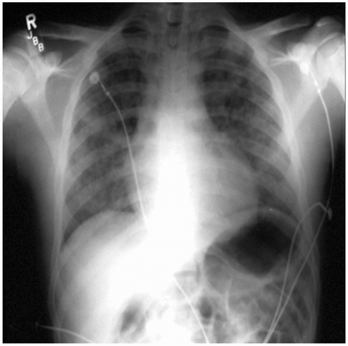
FIGURE 5.1 – Diffuse pulmonary infiltrates consistent with interstitial pulmonary edema following tracheal extubation and transient airway obstruction.


What Is Postobstructive Pulmonary Edema?


▪ HISTORICAL CONSIDERATIONS

In the case under discussion, several questions might be asked—most, if not all of them, after the fact. The questions of utmost importance are, what happened, and was it preventable? Almost certainly this young man sustained postobstructive pulmonary edema, often referred to (incorrectly) as negative-pressure pulmonary edema. The clinical course of this problem has been reported in the published literature for more than 40 years in pediatric patients17,18 and adults,19 and the epidemiology has been reasonably well documented (see Tables 5.1 and 5.2). Could anything have been gleaned from this young man’s history and physical examination? The only “abnormal” finding was a class III Mallampati score, and the value of that routine examination has been subjected to recent criticism.20 Regardless, in this case, the potentially difficult airway or intubation that may have been indicated as a risk did not materialize.


TABLE 5.1 Adult Epidemiology in Postobstructive Pulmonary Edema


Age (years)


37.6 ± 16.6


Range 12-79


Male:Female


1.2:1


Common obstructive events


Laryngospasm (18/32)


Airway tumor (7/32)


Time to onset following obstruction (minutes)


26 ± 39


Range 3-150


Resolution (hours)


30 ± 19


Range 6-72


Data from Lang SA, Duncan PG, Shephard DAE, et al. Pulmonary edema associated with airway obstruction. Can J Anaesth. 1990;37:210.


▪ ETIOLOGY

This condition will be described in some detail, because it is most common to anesthesiology. Other forms of pulmonary edema will be described in Chapter 12. The etiology involves a series of events characterized most commonly by sudden relief of a partial or total obstruction of the airway as, for example, with tracheal intubation (see Table 5.3)—a syndrome most common in anesthesiology. Luke et al.17 reported “… Four patients (ages 3 to 6 years) with severe nasophayrngeal obstruction (had) … cardiorespiratory complications ranging from moderate cardiac enlargement and right ventricular hypertrophy to cor pulmonale and pulmonary edema. … Wide swings in intrathoracic pressure probably played an important role in the etiology of pulmonary edema.”

Capitanio and Kirkpatrick18 noted, “Obstructing lesions of the upper airway should be suspected when the transverse diameter of the heart appears larger during the expiratory phase of respiration as opposed to its size during inspiration. … Acute pulmonary edema without cardiac enlargement may occur in patients with an acute upper airway obstruction.” Oswalt et al.19 described, “Acute fulminating pulmonary edema (that) developed in three patients after acute airway obstruction… (minutes to hours). … The common etiologic factor was vigorous inspiratory effort against a totally obstructed upper airway (and) ventilatory assistance (was required) to maintain oxygenation…”


TABLE 5.2 Pediatric Epidemiology in Postobstructive Pulmonary Edema


Age (years)


3 ± 2.4


Range 1/12-10


Male:Female


2.4:1


Common obstructive events


Supraglottitis (15/45)


Croup (18/45)


Time to onset following obstruction (minutes)


33 ± 66


Range 5-240


Resolution (hours)


42 ± 31


Range 2-96


Data from Lang SA, Duncan PG, Shephard DAE, et al. Pulmonary edema associated with airway obstruction. Can J Anaesth. 1990;37:210.


TABLE 5.3 Etiology of Postobstructive Pulmonary Edema


Relief of obstruction


↓ Airway pressure


↓ Pleural pressure


↑ Venous return and pulmonary artery blood flow


↑ Microvascular pressure


Result: interstitial and alveolar edema


Modified from Kamal RS, Agha S. Acute pulmonary edema. A complication of upper airway obstruction. Anaesthesia. 1984;39:464.


Although one might briefly consider a differential diagnosis—including anaphylactic or anaphylactoid reactions, pulmonary aspiration of gastric contents, some type of latent cardiomyopathy (way down the list)—a history of a brief period of “laryngospasm” during emergence from anesthesia, the benign nature of the clinical course, and the rapid response to minimal therapy—argue for airway obstruction as the causative factor and against anything else.


▪ CLINICAL COURSE

Fortuitously, most cases of this syndrome proceed more or less uneventfully, with rapid recovery being the norm. McConkey21 reported a small series of patients with postextubation pulmonary edema. Of the six individuals studied, all cases were preceded by an episode of laryngospasm; frank hemoptysis occurred in five; one patient was reintubated and ventilated; two patients were admitted to the ICU for face mask CPAP; one patient was managed with CPAP in the PACU; two patients received only oxygen; and all cases resolved fully within 24 hours.


▪ EPIDEMIOLOGY

Published literature suggests that postobstructive pulmonary edema occurs in 20,000 to 30,000 cases annually in the United States and is often unrecognized or misdiagnosed.21 However, the benign nature of this problem is not always seen. Adolph, et al., stated, “Although the vast majority of cases resolve quickly and with minimal problems, death from ARDS and multisystem organ failure has been reported despite relief of the airway obstruction.”21

Postobstructive pulmonary edema is only one of many entities that manifest similar signs and symptoms (see Table 5.4). This similarity makes the historical aspects of a given case extremely important, because the prognosis is considerably better in postobstructive pulmonary edema than, for example, in aspiration of gastric content or other forms of acute respiratory distress syndrome (ARDS). Therapy, for the most part, is simplified, because mechanical ventilation seldom is required, unlike more severe forms of pulmonary edema that are discussed in Chapter 12. Table 5.5 shows the elements that should be considered.


TABLE 5.4 Clinical Presentation of Postobstructive Pulmonary Edema


Respiratory distress


—Tachypnea


—Dyspnea


Paradoxical Breathing


↓ SpO2


Cyanosis (late)


Wheezing, stridor


Pink, frothy secretions (sometimes frank blood)


In summary, postobstructive pulmonary edema probably occurs much more commonly than is generally recognized in the operative and perioperative periods. Its features can easily be mistaken for other conditions such as the pulmonary aspiration of gastric contents, but it differs in that resolution is usually rapid and complete within hours of the inciting episode. Subtle episodes may be detected by pulse oximetry when clinical manifestations are minimal to absent. Prevention clearly is superior to treatment, but the onset is so rapid in some cases that the anesthesia provider may be unaware that any adverse outcome related to airway obstruction has occurred. Therefore, the answer to the second question asked at the beginning of this chapter, “Is the condition preventable?” is often “no”.

Although rare, severe cases leading to death22 have been reported, as has pulmonary hemorrhage.23,24 The incidence of postobstructive pulmonary edema has been suggested to be as high as 0.5% to 1% of all intubated, anesthetized patients. Some cases have resulted from biting and occluding endotracheal tubes and laryngeal mask airways.25,26 They have also occurred in patients for whom there was no evidence of obvious airway obstruction, although relative compromise was likely to be present.


TABLE 5.5 Supportive Therapy of Postobstructive Pulmonary Edema


General supportive measures


Maintenance of patent airway


Oxygen


CPAP


Mechanical ventilation with PEEP (seldom necessary)


Careful monitoring (invasive seldom required)


No furosemide or other diuretics initially


Aggressive fluid administration if necessary to restore depleted intravascular volume in cases of fulminant pulmonary edema


CPAP, continuous positive airway pressure; PEEP, positive-end expiratory pressure.


Is Atelectasis a Major Problem in Perioperative Anesthesia Care?

Atelectasis is a major component of acute respiratory failure and is often the abnormality for which mechanical ventilatory support is perceived to be necessary. It may occur when unopposed elastic recoil of the lungs leads to a decrease in lung volume. Small airways collapse follows, and gas trapped in the alveoli is resorbed into the pulmonary blood (the total partial pressure of gas in the mixed venous blood is always less than atmospheric pressure).27 As the alveoli progressively decrease in volume, a point is reached at which they collapse and become devoid of air. In this state, gas exchange does not occur in the affected alveoli. The clinical manifestations of this condition include increased work of breathing as the affected individual “tries” to reinflate the collapsed alveoli during inspiration; decrease in the ventilation/perfusion [V with dot above]/[Q with dot above] ratio; increase of intrapulmonary shunt ([Q with dot above]sp/[Q with dot above]t) and hypoxemia.

Atelectasis is aided and abetted by a number of factors, many of which are incidental to the administration of a general anesthetic. Included in this list are the breathing of a gas mixture with an increased fraction of inspired oxygen (FIo2) above 0.6; loss of chest wall integrity that normally opposes lung collapse; any condition leading to airway obstruction or collapse; pneumothorax or hemothorax; and intubation of a main stem bronchus (partial or total contralateral lung collapse). In some cases, treatment is relatively straightforward (i.e., repositioning of an endotracheal tube). In others, such as ARDS, it is much more complex.


▪ HISTORICAL CONSIDERATIONS

Concepts involving treatment of atelectasis by the application of mechanical ventilation trace their origins to Bendixen et al.28,29,30 and their investigations of anesthetized patients in the 1960s. They described gradually increasing degrees of hypoxemia in spontaneously breathing patients undergoing surgical procedures and attributed the change to “miliary atelectasis” that occurred as a result of “monotonous” low tidal volume (VT) breathing. Mechanical ventilators were rare in most operating theaters at that time, and patients were often allowed to breathe spontaneously during the administration of general anesthesia. Because of the respiratory depressant effects of many anesthetic agents or adjuvants, the placement of retractors and packing in the abdominal cavity that impaired diaphragmatic excursion, and the lack of airway humidification that contributed to inspissation of secretions, predisposition to atelectasis was common. Although later work showed that other factors, including alterations in position and changes in diaphragmatic mechanics,30 were as important, the concept of low VT as a cause of atelectasis was firmly entrenched.

Only gold members can continue reading. Log In or Register to continue

Stay updated, free articles. Join our Telegram channel

Jul 15, 2016 | Posted by in ANESTHESIA | Comments Off on Respiratory Complications Overview

Full access? Get Clinical Tree

Get Clinical Tree app for offline access