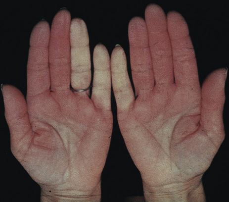
Lin A. Brown Raynaud phenomenon is a reversible vasospastic disorder that affects the blood flow to the digits. When these changes occur in isolation with normal physical examination findings, the disorder is known as primary Raynaud phenomenon. In primary Raynaud phenomenon, there are no associated autoimmune diseases, and rarely are autoantibodies present. Raynaud phenomenon characteristically occurs in women with a mean age of 47 to 53. It is not uncommon and is thought to affect 9% to 10% of women and 5% to 6% of men in the United States.1 Eighty percent of cases of primary Raynaud phenomena occur in non-Latino whites with age younger than 30, positive family history, and symmetric color changes with mild intensity.2 Secondary Raynaud phenomenon is seen in patients who also have an autoimmune disorder, such as progressive systemic sclerosis (scleroderma), systemic lupus erythematosus, dermatomyositis, polymyositis, or mixed connective tissue disease. Secondary Raynaud phenomenon can also be seen as part of the CREST constellation, a subset of scleroderma characterized by calcinosis, Raynaud phenomenon, esophageal dysmotility, sclerodactyly, and telangiectasia. In addition to autoimmune diseases, Raynaud phenomenon has been seen in association with migraine headaches and chest pain.3 Secondary Raynaud phenomenon is often more severe than primary disease and less symmetric in the digits involved and has a greater likelihood of ulcerations and severe ischemic changes. Secondary Raynaud phenomenon can be associated with certain drugs, trauma, hematologic disorders, vibration exposure, frostbite, and atherosclerotic disease. More than 60% of patients who develop Raynaud after age 60 have atherosclerosis as the underlying cause of their disease.2 The vascular endothelium, smooth muscle cells, and nerve terminals form an integrated unit that works in response to elements in the microenvironment to determine the final balance between vasodilation and vasoconstriction. These elements are influenced by a variety of factors, including temperature, physical activity, emotional state, and direct trauma. In Raynaud phenomenon, the blood vessels constrict in response to cold or stress. This may be related to increased activation of the sympathetic nerves, especially the alpha2-adrenergic fibers. In addition, there appears to be an alteration in vascular function on the cellular level, particularly in vascular smooth muscle and the endothelium.4 The resultant disturbance in circulation causes a series of color changes in the skin: white, blanched, or pale as the blood flow is reduced (Fig. 217-1); blue as the affected digit loses oxygen from the decreased blood flow, and red or flushed as blood flow returns. Finally, as the attack subsides and the circulation returns to normal, usual skin color is restored. In the white and blue stages, numbness, tingling, and coldness can be felt. In the red stage, a feeling of warmth, burning, or swelling may be reported. Not infrequently, pain is experienced, especially in secondary Raynaud phenomena. The earlobe, nose, and rarely tongue can be affected as well as the fingers and toes. The vasospasm of Raynaud phenomenon causes classic tricolor changes of first white (pallor), then blue (cyanosis), and then red (reperfusion hyperemia) after the vasospasm ends.1 Episodes can be triggered by cold exposure, rapid changes in ambient temperature, or emotional stress. Attacks can occur in single or multiple digits and can spread to other digits, the other hand, or the feet. Cutaneous vasospasm can be seen at other sites, including ears, nose, face, knees, and nipples.5 Patients can experience pain, numbness, and burning, especially in secondary Raynaud phenomena. Episodes last between 20 minutes and hours. Patients with a history of digital frostbite can subsequently experience Raynaud phenomenon in the affected digits. The diagnosis is based on the presence of two or more color changes with sharp demarcation on the digits. Color photographs can assist in making the diagnosis.6 On examination, the aforementioned classic tricolor changes may be observed with sharp demarcation where the spasm occurs. Submersion of the patient’s hand in ice water is not recommended. Physical examination of the digits can reveal dilated capillary loops at the base of the nail beds as a sign of secondary Raynaud phenomena. Nail fold capillaroscopy is performed by observing the capillary loops in the cuticle adjacent to the nail. Immersion oil or K-Y Jelly is helpful in making the keratin more translucent and therefore accentuating the capillary loops. Magnification can be achieved with an ophthalmoscope or with another magnifying instrument A DermLite can be especially helpful. Microvascular abnormalities such as tortuous or dilated capillary loops and capillary dropout are observed in secondary Raynaud phenomena. Other stigmata of connective tissue disease may be observed on physical examination, such as the sclerotic hidebound skin of scleroderma, rash, mechanic’s hands dermatitis or polymyositis, and arthritis. Tissue breakdown and ulcerations, or pitted scars of former attacks, can also be present. The diagnosis of Raynaud phenomenon is based on a clinical history of the classic tricolor changes. Capillaroscopy of the nail fold allows direct visualization of the capillaries, helping to distinguish primary from secondary Raynaud phenomenon.7 Antinuclear antibody (ANA) positivity is a predictor of progression to or association with a connective tissue disease.8 In particular, the presence of an anticentromere antibody is associated with the development of CREST (the subset of scleroderma described earlier), whereas the anti–Scl-70 antibody is more often seen in scleroderma.9 One source notes that 1% of patients with primary Raynaud phenomenon converted to secondary Raynaud phenomenon per year. However, 47% of patients with primary Raynaud phenomenon who had either ANA positivity or capillary changes progressed, compared with 80% if both were present (ANA and capillary changes).10 Diagnostics Raynaud Phenomenon
Raynaud Phenomenon
Definition and Epidemiology
 Physician consultation is recommended for patients with persistent pallor, coldness, or tissue breakdown in the digits and reduced pulses in the extremities.
Pathophysiology
Clinical Presentation
Physical Examination
Diagnostics
Raynaud Phenomenon
Chapter 217