RASH: NEONATAL
LESLIE CASTELO-SOCCIO, MD, PhD
Rashes are common in the neonatal period and can cause significant parental distress. The ability to distinguish worrisome rashes from those that are benign is of critical importance. To provide a schema for understanding rashes in the neonate, it can be helpful to divide the rashes into categories: pustules, vesicles, patches/plaques, birth marks/hamartomas, and dyspigmentation. Within these categories, there are sign and symptoms that push the clinician to be more or less concerned.
PUSTULAR (NEONATAL ACNE)
Pustular rashes in neonates are common and can be caused by inflammation (such as in erythema toxicum and transient neonatal melanosis) or infections (yeast, bacteria like Staphylococcus aureus, and, rarely, herpes virus [please see vesicular neonatal rashes below for full discussion of herpes simplex]). The goal of recognition is to spare healthy infants with benign pustular eruptions extensive workups and not to miss those with more serious pustular eruptions.
Erythema Toxicum Neonatorum
Erythema toxicum neonatorum (ETN) is usually evident within the first 48 hours of life. Characteristic lesions include erythema, wheals, papules, and pustules (Fig. 64.1). This transient rash resolves spontaneously without sequelae over the course of 1 to 2 weeks. Histologically, ETN shows an abundance of eosinophils. Etiology is unclear. One prospective study of 1,000 neonates suggested that risk factors include higher birth weight, greater gestational age, vaginal delivery, maternal age <30 years, and fewer than two previous pregnancies. Culture and Gram stain looking for eosinophils can help distinguish from bacterial infection. A similarly benign pustular eruption in neonates is transient neonatal pustular melanosis (TNPM), which is usually present at delivery. TNPM is characterized by small pustules (0.3 to 0.5 cm) on a nonerythematous base. These pustules rupture easily, and pigmented macules develop with surrounding collarettes of scales that may persist for weeks to months. The pustules are mostly located over the forehead, neck, and lower back, but occasionally, palms and soles may be involved (Fig. 64.2). No systemic manifestations have been reported.
Staphylococcal Pustulosis
If a neonate presents after 48 hours with new pustules, it is more likely to be caused by infection with staphylococcus or candida. Staphylococcal pustulosis is relatively common and can occur in the setting of infection of the umbilicus or circumcision site. Community-acquired S. aureus is common, so risk factors for acquiring staph in neonatal period include history of staph infection in close contacts. Simple bacterial swabs are the primary diagnostic tool. Pustules on the lower abdomen and in the diaper area are common. Few pustules, in an otherwise healthy neonate, can often be treated with oral and/or topical antibiotics. Providers should look for peeling in the folds of the skin and very red or hot skin because this can be a sign of staphylococcal scalded skin (Fig. 64.3).
Neonatal Candida
Congenital candidiasis usually presents within 12 hours of birth as redness on the affected area and then later with pustules with desquamation. Candida albicans and Candida psiloparis are the most common causes of neonatal candida infections. In full term healthy infants, congenital candida can often be treated topically and is usually a nonworrisome infection. In preterm infants, or other medically complex infants, candida can be invasive and can cause late-onset neonatal sepsis. Therefore, blood cultures, urine cultures, evaluation of the CSF, opthalmologic examination, echocardiogram, renal ultrasound, and systemic antifungal therapy are needed.
Moniliasis/Candidal Diaper Dermatitis
See Chapter 61 Rash: Bacterial and Fungal Infections. Moniliasis is the most characteristic of the diaper rashes (see below for a more detailed discussion of diaper dermatitis). The skin in the diaper area has clusters of erythematous papules and pustules that coalesce into an intensely red confluent rash with sharp borders (Fig. 64.4). Beyond these borders are satellite papules and pustules. At times, the infant has concomitant oral thrush. When the problem is chronic and recurrent, seeding from the gastrointestinal (GI) tract or from a mother with monilial vaginitis should be considered.
On rare occasions, an id reaction occurs. Besides the primary monilial diaper rash lies an antigenic dissemination with involvement of the intertriginous areas and scattered small patches or plaques of scaling erythema on other parts of the skin surface. Generally, C. albicans cannot be cultured from the plaques of the id reaction.
INFANTILE ACROPUSTULOSIS
Infantile acropustolosis is a recurrent, self-limited, vesiculopustular disorder affecting young children (Fig. 64.5). It presents with itchy, deep-seated pustules or vesicles on the palms and soles. Most cases occur after scabies infestation and this condition can wax and wane for years. Scraping for scabies mites should be performed and treatment initiated for scabies if mites are found. Therapy is midpotency topical steroids.
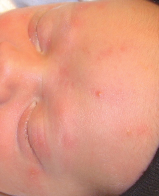
FIGURE 64.1 Infant with papules and pustules of erythema toxicum on the face.
VESICULAR ERUPTIONS
Neonatal herpes infection causes significant morbidity and mortality if not recognized and treated promptly (see Chapter 68 Septic Appearing Infant). Neonatal HSV can be acquired in utero (5%), in the peripartum period (85%) or in the postnatal period (10%). For the last two groupings, the extent of disease can be classified into the following categories: disseminated disease, central nervous system (CNS) disease, or skin, eye, and/or mouth (SEM) disease. A high index of suspicion is necessary in neonates presenting with vesicles or pustules, especially with a negative bacterial culture. About 40% of neonatal herpes is confined to the SEM. Most infections acquired during the peripartum period present between 9 and 11 days of life, though they can be seen earlier or later. Those infants with SEM disease are most easily diagnosed since they usually present with obvious vesicular lesions. HSV that develops in the skin usually begins as papules or vesicles that erode. Often erosions are the only visible lesions. They usually have a red base and are 1 to 3 mm in diameter and can occur as a single unit or in clusters (Fig. 64.6). They appear anywhere on the body but are most commonly seen on the presenting parts such as the head in vertex presentation and buttocks in breach. Scalp probe sites can become sites of primary infection. The poorest outcome is in infants who present with widespread disease involving lungs, liver, adrenal gland, skin, eyes, and mouth. Infants presenting with disseminated herpes disease typically present with symptoms very similar to those associated with bacterial infection. Although the diagnosis may be easily confused, disseminated herpes disease may often be distinguishable from bacterial infection by the presence of vesicular lesions, neonatal hepatitis of unknown etiology, and DIC. Disseminated herpes infection may have a component of CNS involvement and the infant may therefore exhibit symptoms consistent with encephalitis or meningitis. Intrauterine HSV infection can result from primary or recurrent maternal infection. Highest risk occurs when the mother has true primary infection at the time of delivery; the risk for developing neonatal herpes is about 30%. The risk for transmission in infants born to mothers with known genital herpes is less than 1%, which may be related to transfer of maternal HSV IgG antibodies across the placenta.

FIGURE 64.2 Multiple collarets of scale on the leg of a newborn with TNPM.
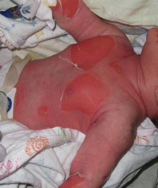
FIGURE 64.3 Skin peeling on the trunk of infant with staphylococcal scalded skin syndrome.
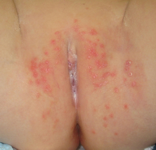
FIGURE 64.4 Infant with candidal diaper dermatitis.

FIGURE 64.5 Acropustulosis of infancy.
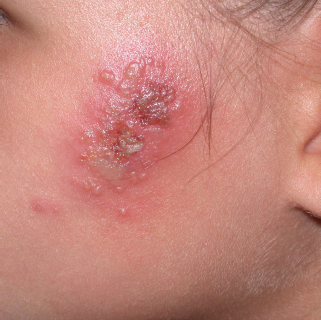
FIGURE 64.6 Clusters of vesicles and erosions on the cheek of a child with HSV.
Since neonatal herpes infection can be seen up until 4 weeks of age, clinicians caring for newborns in clinics, offices, and emergency department must be familiar with the clinical manifestations of this disease process. Once there is suspicion of neonatal HSV, obtain the following samples before starting antiviral therapy:
1. CSF for indices and HSV DNA PCR,
2. swab for viral culture +/– PCR from base of vesicles,
3. swab from the mouth, conjunctiva, nasopharynx and rectum for viral culture +/– PCR, and
4. whole blood for HSV DNA PCR.
Start empiric intravenous acyclovir therapy (60 mg/kg/day divided in 3 doses). For SEM disease, treat for 14 days. For disseminated and CNS diseases, treat for 21 days and then treat with oral acyclovir suppressive therapy for 6 months (900 mg/m2/day divided in 3 doses). Also, for both CNS and disseminated diseases, repeat CSF analysis and CSF HSV PCR before stopping therapy and, if detectable in CSF, continue therapy until negative CSF PCR results. Dose of acyclovir should be weight-adjusted for the duration of therapy. Absolute neutrophil count should be monitored every 2 weeks for the first month and then monthly.
INCONTINENTIA PIGMENTI
Incontinentia pigmenti (IP) is a rare X-linked genodermatosis that affects mainly female neonates (see Chapter 62 Rash: Vesiculobullous). The first manifestation occurs in the early neonatal period and progresses through four stages: vesicular, verruciform, hyperpigmented, and hypopigmented. Clinical features also manifest themselves through changes in the teeth, eyes, hair, CNS, bone structures, skeletal musculature, and immune system. IP is often mistaken for an infectious process (Fig. 64.7). PCR and cultures of the vesicles yield negative results. The clue to this diagnosis is that the vesicles usually occur on the arms and/or legs in a linear pattern that follows the lines of Blaschko. Genetic testing and/or a biopsy can help confirm the diagnosis. Rarely, boys that are XXY or have somatic mosaicism present with IP.

FIGURE 64.7 Linear vesicles in a newborn with the first stage of incontinentia pigmenti.
PATCHES AND PLAQUES
Annular Rashes
Neonatal lupus is an autoimmune disorder caused by the passive transfer of maternal autoantibodies, anti-Ro, anti-La, and, less commonly, anti-ribonucleoprotein (U1-RNP) (Fig. 64.8). The skin and heart are commonly affected, with the most serious complication being third-degree atrioventricular heart block (AVB), which results in fetal and neonatal mortality rates of 15% to 30%. Ten percent of patients experience thrombocytopenia, neutropenia, or anemia, which are usually transient. Neonatal lupus can present as annular, red, scaly patches, most commonly on the head and neck. Rash around the periorbital region should make suspicion very high (Fig. 64.9)
Related posts:


Stay updated, free articles. Join our Telegram channel

Full access? Get Clinical Tree


