RASH: DRUG ERUPTIONS
MELINDA JEN, MD
DRUG ERUPTIONS
The spectrum of cutaneous drug eruptions ranges from the relatively benign, where the medication can be continued if essential, to the severe, where there can be significant morbidity and mortality. Thus, in cases of severe drug reactions, prompt and accurate diagnosis is critical and can be lifesaving. The primary morphology of the eruption helps guide the clinician to a diagnosis. Herein, we summarize the most salient features of the most common drug reactions, with a particular focus upon their primary morphologies.
URTICARIA
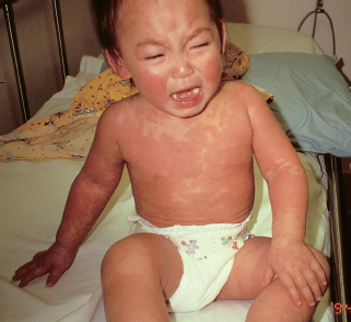
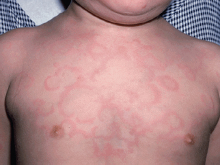
Urticaria (hives, wheals) consists of erythematous, edematous papules and plaques that can coalesce into larger polycyclic, arcuate, and annular plaques (Figs. 63.1 and 63.2). A key diagnostic feature is that individual lesions are transient, resolving within 24 hours, but with new lesions appearing elsewhere. As they resolve, purpuric macules secondary to capillary leak and hyperpigmentation may remain. Pruritus and angioedema, particularly of the eyelids, hands, and feet, are common.
Urticaria results from IgE degranulation of mast cells. Although the most common cause of urticaria is infection, medications can sometimes trigger urticaria. Urticaria typically appears within the first 2 weeks of starting the culprit medication. Cephalosporins, β-lactam antibiotics, sulfonamides, and anticonvulsants are common causes of drug-induced urticaria. Some medications, such as nonsteroidal anti-inflammatory drugs (NSAIDs), may cause urticaria through both immunologic and nonimmunologic pathways (via increased leukotriene synthesis).
Urticaria is often confused for erythema multiforme (EM). The key features differentiating urticaria from EM are morphology, individual lesion duration, symptomology, and distribution. Urticaria can be annular, polycyclic, and arcuate, but does not have the classic target appearance of EM. Additionally, urticaria does not vesiculate, while the central areas of EM lesions may be bullous. As noted above, individual lesions of urticaria last less than 24 hours, while lesions of EM are fixed and take several days to resolve. If symptomatic, urticaria is pruritic while EM may itch or burn. Urticaria can occur anywhere on the body, while EM typically first appears on the hands and feet before progressing centrally.
The treatment for drug-induced urticaria is withdrawal of the causative medication and treatment with both H1 and H2 blocking oral antihistamines for up to 4 to 8 weeks. For cases that persist despite maximum dosing of nonsedating H1 and H2 blocking antihistamines, oral steroids may be beneficial.
MORBILLIFORM
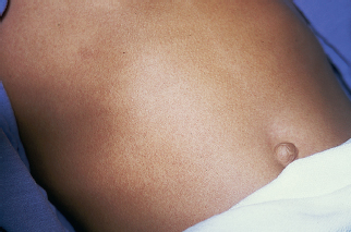
Morbilliform, or measles-like, describes an eruption characterized by both erythematous macules and papules (Fig. 63.3). The term “maculopapular” is often used to describe this eruption, but morbilliform is a more precise description. This is the most common type of drug rash. The eruption typically starts on the trunk before spreading to involve the extremities and face, though the mucous membranes are spared. The eruption can become diffuse and confluent. Pruritus may be present.
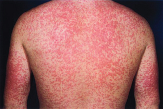
A morbilliform drug eruption typically appears 7 to 14 days after medication initiation, but in a previously sensitized patient it may appear within hours to days of reexposure. Antibiotics, in particular penicillins (Fig. 63.4), cephalosporins, and sulfonamides, as well as antiepileptics are common triggers.
A morbilliform drug eruption can be difficult to clinically distinguish from a viral exanthem. One unique example is that of the morbilliform eruption that may result from antibiotic administration, most commonly amoxicillin, during an Epstein–Barr virus (EBV) infection. Although early studies reported an incidence of 90% or more, a more recent study suggests that the true incidence is closer to 30%. This eruption, however, is not actually a drug hypersensitivity reaction (DHR), but rather is a viral exanthem.
Treatment involves a balance between the severity of the eruption and the importance of the causative medication. The rash is generally self-limited and will resolve within 7 to 14 days of stopping the medication. However, if the rash is mild and the medication is essential, then the medication can be continued with close monitoring. Morbilliform drug eruptions do not progress into more severe drug reactions, however, early on severe drug reactions may mimic a morbilliform drug eruption. Therefore, an uncomplicated morbilliform drug eruption must be distinguished from DHR, which has systemic involvement. If the eruption appears within the first 2 weeks of starting a medication, then it is more likely to be a morbilliform drug eruption. If the eruption is delayed by several weeks, then DHR is more likely. The presence of additional clinical features, such as facial edema and lymphadenopathy, and laboratory findings can also aid in distinguishing the two entities. If needed, topical steroids can help provide symptomatic relief of pruritus.
DRUG HYPERSENSITIVITY REACTION (DHR)/DRUG REACTION WITH EOSINOPHILIA AND SYSTEMIC SYMPTOMS (DRESS)
The cutaneous eruption of DHR, also known as drug reaction with eosinophilia and systemic symptoms (DRESS), is a morbilliform eruption that starts on the face and spreads cephalocaudally. Additional clinical findings and systemic involvement distinguish DHR from a skin-limited morbilliform drug eruption. Facial edema is present in approximately 76%, fever in 90%, and lymphadenopathy in 54% of patients with DHR. In half of patients, there can be mild mucosal involvement, more commonly the oral mucosa. Systemic involvement commonly manifests with eosinophilia (95%) or atypical lymphocytosis (67%) on complete blood count. Liver involvement is seen in 75% of patients, and often presents as elevation of liver transaminases. In cases where patients have a prolonged clinical course or when they appear systemically ill, echocardiogram, renal function tests, and coagulation profiles should be checked for cardiac, renal, and severe hepatic involvement. Thyroid involvement is usually delayed in onset, so thyroid functions should be followed for 2 to 3 months after the DHR.

FIGURE 63.1 Widespread transient erythematous edematous papules and plaques. (From Fleisher GR, Ludwig S, Baskin MN. Atlas of pediatric emergency medicine. Philadelphia, PA: Lippincott Williams & Wilkins, 2004.)

FIGURE 63.2 Urticaria often appears as annular or polycyclic plaques with central clearing or purpura. (From Burkhart C, Morrell D, Goldsmith LA, et al. VisualDx: essential pediatric dermatology. Philadelphia, PA: Lippincott Williams & Wilkins, 2009.)

FIGURE 63.3 A morbilliform eruption presents with erythematous macules and papules on the trunk before spreading to the rest of the body. (Courtesy of George A. Datto, III, MD.)
DHR is a delayed-type hypersensitivity reaction that occurs 2 to 6 weeks, with an average of 22 days, after starting a medication. Antiepileptics (carbamazepine, phenytoin, phenobarbital, lamotrigine) are the most common cause of DHR. Other medications that commonly cause DHR are trimethoprim-sulfamethoxazole, minocycline, dapsone, sulfasalazine, and abacavir. There appears to be a genetic susceptibility for developing DHR to certain medications. For example, HLA-A*31:01 has been showed to be associated with an increased risk of DHR following exposure to carbamazepine in children. Other associations with DHR include human herpesvirus-6 (HHV6), in which reactivation has been detected in cases of DHR. Though the exact role of HHV6 in the pathogenesis of DHR is still unclear, some evidence suggests that HHV6 reactivation creates immune dysregulation that causes DHR, while others suggest that DHR-induced immune dysregulation allows for HHV6 reactivation.

FIGURE 63.4 This morbilliform eruption presented several days after starting ampicillin therapy. (From Elder DE, Elenitsas R, Rubin AI, et al. Atlas and synopsis of lever’s histopathology of the skin. 3rd ed. Philadelphia, PA: Lippincott Williams & Wilkins, 2012.)
Stay updated, free articles. Join our Telegram channel

Full access? Get Clinical Tree


