RASH: BACTERIAL AND FUNGAL INFECTIONS
JAMES R. TREAT, MD
BACTERIAL INFECTIONS
Pustular Eruptions
Folliculitis
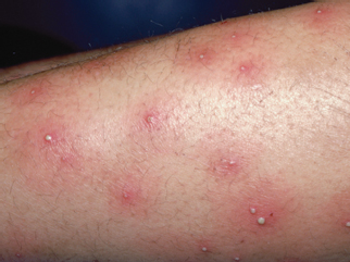
Folliculitis presents with pustules and red papules that are centered around hair follicles due to bacterial invasion of the follicle (Fig. 61.1). The infection is sometimes itchy but can be painful. When excoriated, the pustules may be unroofed and eroded, or crusted papules may predominate. Folliculitis can be widespread but is often concentrated in hair-bearing areas. Cultures of pustules prove the diagnosis, establish bacterial sensitivities, and guide therapy. Folliculitis is most commonly caused by Staphylococcus aureus (SA). Group A Streptococcus (GAS) causes folliculitis as well but may present with intermixed pustules, vesicles, and erosions.
Pseudomonas aeruginosa can survive in recirculated water and causes “hot tub” folliculitis. Pseudomonas infections from hot tubs or other recirculating water often present under the clothing where the bacteria get trapped. Therapy with antibacterial washes and topical antibiotics directed at P. aeruginosa, such as silver sulfadiazine, bacitracin/polymyxin B, gentamicin or neomycin, are often sufficient. For more widespread or symptomatic infections or those in immunocompromised hosts, systemic therapy with ciprofloxacin (off-label) should be considered.
Gram-negative folliculitis can be confused with acne; it can occur on the face and presents in adolescents with pustules, especially around the mouth. The differential diagnosis of bacterial folliculitis includes other nonbacterial causes of folliculitis such as pityrosporum yeast infections, especially on the upper trunk and neck of adolescents, or miliaria (heat rash) in patients who are sweating. Patients who are immunodeficient can have folliculitis due to gram-negative bacteria, disseminated yeast, and mold infections. Performing a swab culture of the pus expressed from an intact pustule establishes the diagnosis and guide therapy.
It is very important to recognize that vesicular eruption of herpes simplex virus (HSV) or varicella zoster virus (VZV) can also look pustular if the eruption has been present for a few days. Neutrophils infiltrate the vesicles and make them look pustular. Therefore, in neonates with pustular eruptions, and any child with recurrent localized areas of grouped pustules, HSV should be considered.
Disseminated Gonococcal Infections (See also Chapter 102 Infectious Disease Emergencies)
Localized genitourinary or oral infection with Neisseria gonorrhoeae can rarely disseminate to the skin, presenting with erythematous papules, petechiae, or vesicle-pustules on a hemorrhagic base. These cutaneous lesions usually develop on the trunk but may occur anywhere on the extremities. Disseminated N. gonorrhoeae should be considered in sexually active or sexually abused children, especially if the partner has a history of vaginal or penile discharge.
Diagnosis can be made by culture of vesiculopustular skin lesions, blood culture, or positive culture of oral or genital sites. Gram stains of pustules show gram-negative diplococci and can help support the clinical diagnosis, although Neisseria meningitidis may have the same appearance on Gram stain. Based on resistance patterns, recommended current therapy is ceftriaxone until clinical improvement is seen, at which point it can be changed to an oral antibiotic, such as cefixime, ciprofloxacin, ofloxacin, or levofloxacin, for a total of a 7-day course. Quinolones should not be used for infections in men who have intercourse with men or in those with a history of recent foreign travel or partners’ travel, or infections acquired in other areas with increased resistance. Concomitant sexually transmitted diseases should be tested for and treated empirically.
Furunculosis
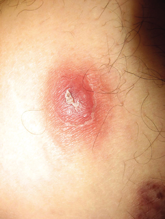
Furunculosis is an acute purulent abscess extending from the dermis into the subcutis. Furuncles manifest as painful red or purple fluctuant nodules with or without a pustule on top (Fig. 61.2). Furunculosis is most commonly caused by SA and can be methicillin sensitive or resistant (MRSA). Diagnosis is clinical but can be aided by ultrasound if it is unclear if there is fluctuance. Therapy is with incision and drainage and adding antibiotic coverage is controversial. The literature suggests antibiotics are not necessary for simple abscesses except in young children unless there is associated cellulitis, the lesion has failed incision and drainage previously, the patient is immunosuppressed or showing signs of sepsis, or the lesion is particularly difficult to drain. Cultures can be sent from the purulent drainage in order to confirm the diagnosis and measure the antibiotic sensitivities. Empiric therapy should be guided by local resistance patterns but is usually with clindamycin, trimethoprim-sulfamethoxazole, or doxycycline (if age appropriate) in order to cover for MRSA.
Recurrence of furunculosis is common. Reinfection from the patient’s local environment can come from sources such as close contacts, pets, athletic equipment, or stuffed animals. Reinfection may occur because of reinoculation from the patient’s own nares. Decolonization is challenging but nasal decolonization with intranasal mupirocin two times daily for 5 days for the patient and any close contacts can be effective. Four percent chlorhexidine washes or dilute sodium hypochlorite (¼ cup in 20 to 40 gallons of water for 15 minutes) baths can be used to decolonize the skin but caution in young children to assure they do not ingest or get the cleanser on the mucosa.

FIGURE 61.1 Folliculitis. (From Burkhart C, Morrell D, Goldsmith LA, et al. Essential pediatric dermatology. Philadelphia, PA: Lippincott Williams & Wilkins, 2009.)
Bullous Impetigo
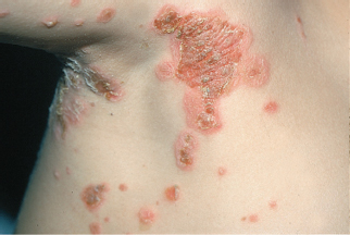
Bullous impetigo is caused by a localized staphylococcal infection that produces an exfoliative toxin that cleaves the skin connection desmoglein 1 (DGS1). This allows fluid to build up within the epidermis and forms bullae (Fig. 61.3). When the bullae rupture, the roof of the bulla and the fluid dries to the skin giving the classic “honey-colored” crusting. SA colonization is most common in the nares and perianal areas and thus impetigo is more common on the face and perineum. Culture of the blister fluid will yield the pathogen and establish sensitivities for therapy. Localized bullous impetigo can often be treated with topical antibiotics such as mupirocin, bacitracin, or retapamulin. More widespread or severe infections or those in immunosuppressed hosts or neonates should be treated systemically.

FIGURE 61.2 Furuncle. Note pustule with surrounding erythema and induration. (Image courtesy of Lee R. In: Elder DE, ed. Lever’s histopathology of the skin. 11th ed. Philadelphia, PA: Wolters Kluwer, 2014.)

FIGURE 61.3 Bullous impetigo. (From Goodheart HP. Goodheart’s photoguide of common skin disorders. 2nd ed. Philadelphia, PA: Lippincott Williams & Wilkins, 2003.)
Staphylococcal-Scalded Skin Syndrome
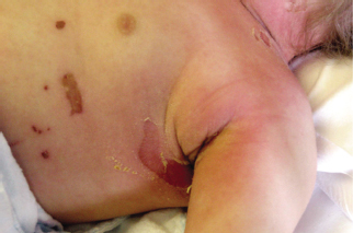
Staphylococcal-scalded skin syndrome (SSSS) is a severe infection resulting from dissemination of the exfoliative toxin produced by SA. SSSS is most common in children under 5 years of age and can present in neonates. The clinical appearance is diffuse redness that parents commonly liken to a sunburn. The skin then peels most characteristically around the mouth, nose, and eyes. Although the crusting and peeling is most prominent in these periorificial areas and can be exuberant, the actual conjunctiva and oral mucosa are not affected. Peeling is also typically prominent in the neck, axillary, and inguinal folds (Fig. 61.4). If the red skin is rubbed, a blister can often be induced (Nikolsky sign). The main clinical differential is Stevens–Johnson syndrome (SJS), which by definition must affect two mucous membranes. In the mucosa, desmoglein 3 is more important in keratinocyte cell–cell adhesion than DSG1. The exfoliative toxin only targets DSG1 and so the mucosa is spared in SSSS. Therefore, the most reliable way of differentiating SSSS from SJS is the lack of mucosal involvement in SSSS. Kawasaki can also cause peeling skin, especially on the hands and feet, but the peeling occurs at least 10 to 14 days after the initial febrile episode, while the exfoliation in SSSS occurs within the first few days of the onset of the illness.

FIGURE 61.4 Staphylococcal-scalded skin syndrome. Note the scalded appearance of the skin under the ruptured bullae of the chest and axilla in this child with staphylococcal-scalded skin syndrome. (From Lippincott Nursing Assessment. February 2014. Philadelphia, PA: Wolters Kluwer, 2014.)
The primary site of the staphylococcal infection is often unknown but recent surgeries (umbilical cord or circumcision in neonates) or other breaks in the skin should be evaluated and cultured. If no clear source of infection can be found, the nares and anus and most heavily crusted areas should be cultured in order to establish antibiotic sensitivities of the SA that is colonizing the child. Therapy for SSSS is with systemic antistaphylococcal antibiotics, typically oxacillin or other beta lactamase–resistant antibiotics. Clindamycin is often added because it inhibits bacterial ribosomal function, thus decreasing toxin formation. In critically ill patients vancomycin should be considered in case the infection is being caused by a resistant staphylococcal strain.
Ecthyma
Ecthyma is a skin infection with loss of the top layers of the skin caused by necrosis. The most common cause of ecthyma in children is GAS and typically presents with painful crusts and erosions (Fig. 61.5)
Stay updated, free articles. Join our Telegram channel

Full access? Get Clinical Tree


