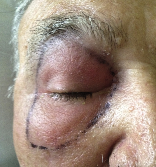
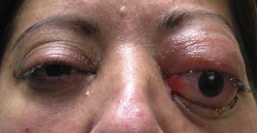
Nathan Blessing, James T. Banta Periorbital infections are classically divided anatomically into whether the findings are isolated anterior to the orbital septum (preseptal cellulitis) or involve structures posterior to the orbital septum (orbital cellulitis). The distinction is critical because infection of the orbit with extension to the orbital apex and beyond can lead to rapid blindness and have potentially fatal consequences. Both disease entities are more common in children, with preseptal infection occurring much more frequently than orbital infection, but individuals of all ages can be affected.1,2 Males are disproportionately affected compared with females at a ratio of 2:1, and the mean age is 7 years in both boys and girls.3 Infection arises from three principle sources: local spread from adjacent structures such as the sinuses and lacrimal system, inoculation after local trauma such as skin lacerations or bug bites, and bacteremic spread from remote foci including the upper respiratory system, middle ear, and heart. As previously mentioned, cellulitis may exist anterior to the orbital septum, with the septum acting as a natural barrier to the passage of microorganisms; however, an initial preseptal cellulitis may spread to involve the orbit as well. There are several anatomic factors that predispose the orbit to infection. Veins in the midface are valveless, permitting the direct posterior spread of infectious organisms into structures such as sinus cavities. The paranasal sinuses surround the orbit inferiorly, medially, and superiorly, and the ethmoid sinus is separated from the orbit by the lamina papyracea, meaning “of paper” because the bone is paper-thin. Unsurprisingly, local spread from ethmoid sinusitis is the most common cause of orbital cellulitis in all age groups, and more than 90% of all cases of orbital cellulitis occur as a secondary extension of acute or chronic bacterial sinusitis.1,4 Causative organisms are usually Streptococcus species or Staphylococcus aureus (including methicillin-resistant S. aureus [MRSA]), but Haemophilus influenzae (less common because of vaccination against H. influenzae type b), Moraxella catarrhalis, and the anaerobic bacteria of the upper respiratory tract may also be implicated.5 In children, the majority of culture results are typically positive for staphylococci or streptococci, with the remainder being polymicrobial. Patients over the age of 15 are more likely to have polymicrobial infections. In immunocompromised or diabetic patients, a fungal cause, such as Aspergillus or Mucor, should be considered.1 Careful physical examination will elucidate key information regarding whether an infection is located anterior or posterior to the orbital septum and the severity of the infection. Preseptal cellulitis typically manifests with eyelid edema, warmth, and erythema that may be severe (Fig. 77-1). The eye is typically spared, without conjunctival injection or chemosis. In contrast, orbital cellulitis causes axial proptosis, lid swelling, conjunctival chemosis and injection, elevated intraocular pressure, and pain or restriction with eye movement (Fig. 77-2).4 The patient may report decreased visual acuity and diplopia. Decreased visual acuity and an afferent pupillary defect (see Chapter 70) are suggestive of optic nerve compromise, one of the most feared complications of orbital cellulitis. Patients with both preseptal and postseptal infections may be febrile, but systemic malaise is typically associated with postseptal disease. A history of sinusitis, local cutaneous infection, trauma, or dental infection is common. The condition is typically unilateral. A history of neck stiffness or mental status change is worrisome for underlying meningitis. If trauma is implicated, the patient usually is seen 48 to 72 hours after the inciting event. However, in the presence of a retained foreign body, the clinical presentation of cellulitis can occur weeks to months after the injury.5 A thorough examination should include an evaluation of the patient’s vital signs, mental status, neck flexibility, visual acuity (including color vision), pupillary response, and extraocular muscle function (cranial nerves III, IV, XI) as well as any pain elicited with extraocular movements. If possible, intraocular pressure should be measured because venous congestion from orbital inflammation may lead to elevated pressures. The globe and ocular adnexa should be inspected for swelling, redness, focal tenderness, hypoesthesia, and fluctuance or drainage. Close inspection should be undertaken to rule out trauma, foreign bodies, or a focal source of infection such as dacryocystitis or eyelid abscess. In diabetic or immunocompromised patients a black eschar located in the nasal cavity or hard palate may herald underlying fungal infection.
Preseptal and Orbital Cellulitis
Definition and Epidemiology
Pathophysiology
Clinical Presentation and Physical Examination
Related posts:


Stay updated, free articles. Join our Telegram channel

Full access? Get Clinical Tree


